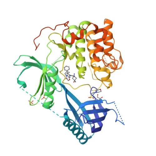
Phosphorylated Antibodies
Phospho-AKT2 specific-S474 Rabbit pAb - P31751
Item Number : CM0001731
Price varies based on specs and customizations
- Application
- WB, ELISA
- Cross Reactivity
- Human
- Protein Weight
- 56kDa

Shipping:
Contact us to get shipping details Enjoy On-time Dispatch Guarantee.
Why Choose Us
Easy ordering process, quality products, and dedicated support for your business success.
Core Product Specifications and Parameters
| Parameter | Value |
|---|---|
| Product Name | Phospho-AKT2 specific-S474 Rabbit pAb |
| Remarks/Alias | PKBB; PRKBB; HIHGHH; PKBBETA; RAC-BETA; Phospho-AKT2 specific-S474 |
| Species | Human |
| GeneID (human) | 208 |
| GeneID | 208 |
| Immunogen | Synthetic peptide |
| Source | Rabbit |
| Category | Phosphorylated Antibodies |
| Application | WB, ELISA |
| Cross Reactivity | Human |
| SWISS | P31751 |
| Protein Weight | 56kDa |
| Shipping | Ice bag |
Biological Background: AKT2 Function and Localization
- AKT2 (RAC-beta serine/threonine-protein kinase, PKB beta) is a member of the AKT kinase family that mediates downstream effects of PI3K signaling, regulating metabolism, proliferation, survival, and growth.
- AKT2 is a critical regulator of insulin-stimulated glucose uptake and glycogen synthesis. It promotes GLUT4 translocation via C2CD5 phosphorylation and inhibits GSK3A/B.
- AKT2 supports cell survival by inactivating pro-apoptotic MAP3K5/ASK1 and by activating CREB1-dependent transcription of BCL2 and MCL1.
- AKT2 is required for SPATA13-mediated regulation of cell migration and adhesion, and may participate in placental development. Related references: PMID:21432781 PMID:21620960
- In response to lysophosphatidic acid, AKT2 suppresses ciliogenesis by phosphorylating WDR44 and RAB3IP. Related references: PMID:31204173
- AKT2 phosphorylates PKP1, enhancing its interaction with 14-3-3 and its nuclear translocation, contributing to reduced keratinocyte adhesion. Related references: PMID:23444369
- AKT2 localizes to the cytoplasm, nucleus, cell membrane, and early endosomes. It is widely expressed, with documented expression in myoblasts. Related references: PMID:17565718
Experimental Guidance and Technical Tips
- The antibody was produced against a synthetic phosphorylated peptide corresponding to Ser474 of human AKT2. To confirm phosphorylation specificity, consider using phosphatase-treated lysates or peptide competition assays.
- For Western blotting, insulin-stimulated cell lysates (e.g., HepG2, 3T3-L1 differentiated adipocytes) may serve as positive controls. A band near 56 kDa is expected.
- In ELISA applications, the phosphorylated immunogen peptide can be used as the coating antigen; cross-reactivity with the non-phosphorylated form should be assessed.
- Since cross-reactivity is currently validated only for human AKT2, verify reactivity in other species if needed.
CamelBio: Your One-Stop Sourcing Bridge
CamelBio provides diagnostic manufacturers, researchers, and laboratories with comprehensive access to high-quality IVD raw materials, technical support, and supply chain solutions. This AKT2 phospho-antibody fits within our portfolio of kinase signaling and metabolism-related reagents, enabling development of assays for diabetes, cancer, and cell survival studies. From validated antibody pairs to bulk ancillary reagents and custom sourcing of rare targets, CamelBio streamlines procurement from concept to clinic.
REQUEST A QUOTE
Our professional team will reply to you within one business day. Please feel free to contact us!