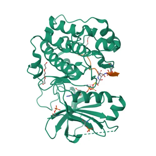
Phosphorylated Antibodies
Phospho-AKT1-T308 Rabbit pAb - P31749
Item Number : CM0001290
Price varies based on specs and customizations
- Application
- WB, ELISA
- Cross Reactivity
- Human, Mouse, Rat
- Protein Weight
- 56kDa

Shipping:
Contact us to get shipping details Enjoy On-time Dispatch Guarantee.
Why Choose Us
Easy ordering process, quality products, and dedicated support for your business success.
Core Product Specifications and Parameters
| Parameter | Value |
|---|---|
| Product Name | Phospho-AKT1-T308 Rabbit pAb |
| Remarks/Alias | AKT; PKB; RAC; PRKBA; PKB-ALPHA; RAC-ALPHA; Phospho-AKT1-T308 |
| Species | Human |
| GeneID (Human) | 207 |
| GeneID | 207 |
| Immunogen | Synthetic peptide phosphorylated around T308 of human Akt1 |
| Source | Rabbit |
| Category | Phosphorylated Antibodies |
| Application | WB, ELISA |
| Cross Reactivity | Human, Mouse, Rat |
| SWISS | P31749 |
| Protein Weight | 56kDa |
| Shipping | Ice bag |
Biological Background: AKT1 Function and Localization
-
AKT1 is a member of the AKT serine/threonine kinase family that mediates diverse cellular processes, including metabolism, proliferation, survival, growth, and angiogenesis by phosphorylating numerous downstream targets. Related references: PMID:11882383 PMID:15526160 PMID:15861136 PMID:21432781
-
It plays a central role in insulin signaling by promoting glucose uptake via SLC2A4/GLUT4 translocation and regulating glycogen synthesis through inhibitory phosphorylation of GSK3A (Ser-21) and GSK3B (Ser-9).
-
AKT1 promotes protein synthesis and cell growth by phosphorylating TSC2 at Ser-939 and Thr-1462, thereby activating the mTORC1 pathway. Related references: PMID:12150915 PMID:12172553
-
It regulates cell survival by inhibiting pro-apoptotic factors: phosphorylating MAP3K5/ASK1 at Ser-83 to suppress oxidative stress-induced apoptosis and phosphorylating BAD to inactivate its pro-apoptotic function. Related references: PMID:11154276 PMID:10926925
-
AKT1 phosphorylates FOXO transcription factors (FOXO1 at Thr-24, Ser-256, Ser-319; FOXO3 and FOXO4 at equivalent sites), leading to their cytoplasmic retention and inactivation, which promotes cell proliferation. Related references: PMID:10358075
-
Subcellularly, AKT1 localizes to the cytoplasm, nucleus, cell membrane, and mitochondrial intermembrane space, consistent with its multifaceted signaling roles.
-
AKT1 is widely expressed in human tissues, with protein levels increased in prostate and breast cancers, particularly the Tyr-176 phosphorylated form showing elevated expression during breast cancer progression.
Experimental Guidance and Technical Tips
- For western blotting, consider using insulin-stimulated or growth factor-treated cell lysates as positive controls, and always include phosphatase inhibitors during sample preparation to preserve phosphorylation at Thr308.
- Validate specificity by comparing signal with a total AKT1 antibody, and test the antibody in the relevant model system (human, mouse, or rat) to confirm expected band at ~56 kDa.
- For ELISA applications, verify detection of the phosphorylated peptide versus the non-phosphorylated counterpart, and optimize coating concentrations and antibody dilutions empirically.
CamelBio: Your One-Stop Sourcing Bridge
CamelBio offers a comprehensive portfolio of IVD raw materials, including validated antibody pairs, optimized monoclonal and polyclonal antibodies, and bulk ancillary reagents, to accelerate your AKT1-related signaling and oncology research. We also provide rare target raw-material sourcing and technical consulting to support your entire assay development journey—from concept to commercialization.
REQUEST A QUOTE
Our professional team will reply to you within one business day. Please feel free to contact us!