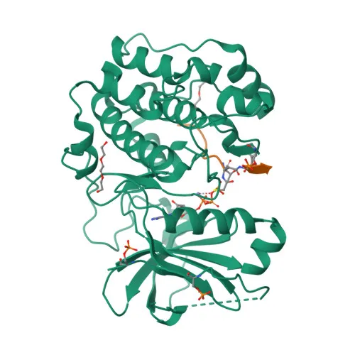
Phosphorylated Antibodies
Phospho-AKT1-S473 Rabbit Polyclonal Antibody for WB, IHC-P, ELISA - P31749
Item Number : CM0001804
Price varies based on specs and customizations
- Application
- WB, IHC-P, ELISA
- Cross Reactivity
- Human, Mouse, Rat
- Protein Weight
- 56kDa

Shipping:
Contact us to get shipping details Enjoy On-time Dispatch Guarantee.
Why Choose Us
Easy ordering process, quality products, and dedicated support for your business success.
Easy Process
Quality Assured
Dedicated Support
Core Product Specifications and Parameters
| Parameter | Value |
|---|---|
| Product Name | Phospho-AKT1-S473 Rabbit pAb |
| Remarks/Alias | AKT; PKB; RAC; PRKBA; PKB-ALPHA; RAC-ALPHA; Phospho-AKT1-S473 |
| Species | Human |
| Gene ID (Human) | 207 |
| Gene ID | 207 |
| Immunogen | Synthetic phosphorylated peptide around S473 of human Akt1 (NP_005154.2) |
| Source | Rabbit |
| Category | Phosphorylated Antibodies |
| Application | WB, IHC-P, ELISA |
| Cross Reactivity | Human, Mouse, Rat |
| SWISS | P31749 |
| Protein Weight | 56kDa |
| Shipping | Ice bag |
Biological Background: AKT1 Function and Localization
- AKT1 is one of three closely related serine/threonine-protein kinases (AKT1, AKT2, AKT3) that regulate metabolism, proliferation, cell survival, growth and angiogenesis. Related references: PMID:11882383 PMID:15526160 PMID:15861136
- AKT1 phosphorylates TBC1D4, triggering binding to 14-3-3 proteins and promoting insulin-stimulated glucose transport into cells. Related references: PMID:11994271
- It activates mTORC1 signaling by phosphorylating TSC2, leading to enhanced protein synthesis and cell growth. Related references: PMID:12150915 PMID:12172553
- AKT1 promotes cell survival by phosphorylating MAP3K5/ASK1, which reduces oxidative stress-induced apoptosis. Related references: PMID:11154276
- It phosphorylates FOXO transcription factors, leading to their cytoplasmic retention and inhibition of pro-apoptotic and cell-cycle arrest genes. Related references: PMID:10358075
- AKT1 positively regulates NF-kappa-B-dependent transcription and CREB1 activity, driving expression of pro-survival genes like BCL2 and MCL1. Related references: PMID:9829964
- The protein localizes to the cytoplasm, nucleus, cell membrane and mitochondrial intermembrane space, reflecting its diverse signaling roles.
- AKT1 expression increases during prostate and breast cancer progression; the Tyr-176 phosphorylated form is elevated in invasive and metastatic stages.
Experimental Guidance and Technical Tips
- The immunogen is a synthetic phosphorylated peptide around S473. To ensure detection of only phosphorylated AKT1, consider using phosphatase inhibitors during sample preparation and including non-phosphorylated peptide blocking controls.
- For Western blot, the predicted molecular weight is 56 kDa. Validate the antibody using lysates from stimulated vs. unstimulated cells.
- In IHC-P, optimize antigen retrieval and include non-phospho peptide blocking to confirm phospho-specificity.
- Cross-reactivity with mouse and rat is reported; however, validate performance in your specific tissue/cell systems.
CamelBio: Your One-Stop Sourcing Bridge
- CamelBio offers one-stop sourcing of IVD raw materials, supporting diagnostic manufacturers and research labs with reliable access to validated antibody pairs, monoclonal and polyclonal antibodies, and bulk ancillary reagents for AKT1 signaling research. Whether you need phospho-specific antibodies, matched detection pairs, or custom rare-target sourcing, CamelBio streamlines your supply chain from concept to clinic.
REQUEST A QUOTE
Our professional team will reply to you within one business day. Please feel free to contact us!