Phosphorylated Antibodies
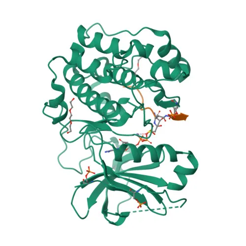
Phospho-Akt-Y315 Rabbit pAb for WB, IHC-P, IF/ICC, ELISA - P31749 / P31751 / Q9Y243
Item Number : CM0001715
Price varies based on specs and customizations
- Application
- WB, IHC-P, IF/ICC, ELISA
- Cross Reactivity
- Human, Mouse, Rat
- Protein Weight
- 48kDa/55kDa/51kDa/54kDa

Shipping:
Contact us to get shipping details Enjoy On-time Dispatch Guarantee.
Why Choose Us
Easy ordering process, quality products, and dedicated support for your business success.
Core Product Specifications and Parameters
| Parameter | Value |
|---|---|
| Product Name | Phospho-Akt-Y315 Rabbit pAb |
| Remarks/Alias | AKT1/AKT2/AKT3; Phospho-Akt-Y315 |
| Species | Human |
| Gene ID (Human) | 207/208/10000 |
| Gene ID | 207/208/10000 |
| Immunogen | Synthetic peptide - A synthetic phosphorylated peptide around Y315 of human AKT1 (NP_001014431.1) |
| Source | Rabbit |
| Category | Phosphorylated Antibodies |
| Application | WB, IHC-P, IF/ICC, ELISA |
| Cross Reactivity | Human, Mouse, Rat |
| SWISS | P31749/P31751/Q9Y243 |
| Protein Weight | 48kDa/55kDa/51kDa/54kDa |
| Shipping | Ice bag |
Biological Background: Akt Function and Localization
-
AKT1 is one of three closely related serine/threonine-protein kinases (AKT1, AKT2, AKT3) that regulate metabolism, proliferation, cell survival, growth, and angiogenesis.
Related references: PMID:11882383, PMID:15526160, PMID:15861136 -
AKT phosphorylates a wide range of downstream substrates on serine/threonine residues, with over 100 reported candidates, though isoform specificity is often unclear.
Related references: PMID:11882383, PMID:15526160, PMID:21432781 -
Key substrates include TSC2, leading to mTORC1 activation and protein synthesis, and FOXO transcription factors, causing 14-3-3 binding and cytoplasmic sequestration.
Related references: PMID:12150915, PMID:12172553, PMID:10358075 -
AKT mediates insulin-induced glucose uptake via phosphorylation of TBC1D4, which promotes binding of 14-3-3 proteins essential for insulin-stimulated glucose transport.
Related references: PMID:11994271 -
Subcellular localization includes cytoplasm, nucleus, cell membrane, and mitochondrial intermembrane space, reflecting its involvement in diverse signaling pathways.
-
AKT is expressed in all human cell types, with increased expression and phosphorylation (e.g., Tyr-176) observed in prostate and breast cancers, linking it to tumor progression.
Experimental Guidance and Technical Tips
- This antibody was raised against a phospho-peptide corresponding to Y315 of human AKT1; consider validating specificity by comparing signals in phosphatase-treated versus untreated samples.
- For Western blot (WB), expected protein weights are approximately 48 kDa (AKT1), 51 kDa (AKT2), and 54 kDa (AKT3); a band around 48–55 kDa may be observed.
- Since the immunogen sequence is conserved, cross-reactivity with human, mouse, and rat Akt is expected; verify in the relevant sample system.
- For IHC-P and IF/ICC, titrate the antibody and include appropriate controls (e.g., peptide blocking) to confirm phospho-specific staining.
CamelBio: Your One-Stop Sourcing Bridge
CamelBio offers diagnostic manufacturers, laboratories, and research institutes one-stop access to IVD raw materials, technical services, and consulting from concept to clinic. This phospho-Akt-Y315 rabbit polyclonal antibody supports your studies on Akt signaling in cancer, metabolism, and cell survival. Beyond individual antibodies, we supply validated antibody pairs, optimized bulk ancillary reagents, and can source rare-target raw materials to streamline your development pipeline.
REQUEST A QUOTE
Our professional team will reply to you within one business day. Please feel free to contact us!