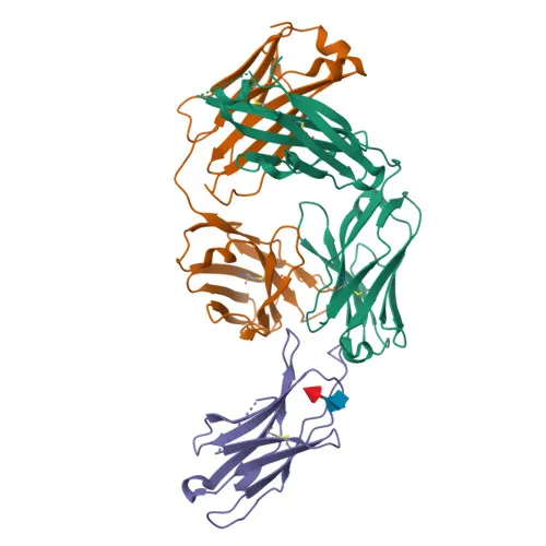
Monoclonal Antibodies
PerCP/Cyanine5.5 Rabbit anti-Human/Monkey PD-1/CD279 Monoclonal Antibody for Flow Cytometry - Q15116
Item Number : CM0011565
Price varies based on specs and customizations
- Application
- FC
- Cross Reactivity
- Human, Cynomolgus
- Protein Weight
- 32 kDa

Shipping:
Contact us to get shipping details Enjoy On-time Dispatch Guarantee.
Why Choose Us
Easy ordering process, quality products, and dedicated support for your business success.
Core Product Specifications and Parameters
| Parameter | Value |
|---|---|
| Product Name | PerCP/Cyanine5.5 Rabbit anti-Human/Monkey PD-1/CD279 mAb |
| Remarks/Alias | PD1; PD-1; CD279; SLEB2; hPD-1; hPD-l; hSLE1 |
| Species | Human |
| Gene ID (Human) | 5133 |
| Gene ID | 5133 |
| Immunogen | Recombinant protein (Recombinant fusion protein containing a sequence corresponding to amino acids 25-168 of human PD-1/CD279 (NP_005009.2)) |
| Source | Rabbit |
| Category | Monoclonal Antibodies |
| Application | FC |
| Cross Reactivity | Human, Cynomolgus |
| SWISS | Q15116 |
| Protein Weight | 32 kDa |
| Shipping | Ice bag |
Biological Background: PD-1 Function and Localization
-
Programmed cell death protein 1 (PD-1, CD279) is an inhibitory receptor expressed on antigen-activated T-cells that plays a critical role in the induction and maintenance of immune tolerance to self. Related references: PMID:21276005 PMID:31754127 PMID:32184441
-
PD-1 delivers inhibitory signals upon binding to its ligands PD-L1 (CD274) and PD-L2 (CD273), leading to attenuation of T-cell receptor (TCR) signaling and suppression of T-cell activation. Related references: PMID:21276005 PMID:26602187
-
Following ligand engagement, PD-1 is phosphorylated within its ITSM motif, recruiting the tyrosine phosphatase SHP-2 (PTPN11), which dephosphorylates key proximal TCR signaling molecules such as ZAP70, PKCθ, and CD3ζ, thereby directly inhibiting T-cell activation. Related references: PMID:32184441
-
Tumors exploit the PD-1/PD-L1 inhibitory axis to evade immune surveillance; interaction with PD-L1 on tumor cells inhibits cytotoxic T lymphocyte (CTL) effector functions, promoting immune escape. Related references: PMID:28951311
-
Blockade of the PD-1 pathway reverses T-cell exhaustion, restores antitumor immunity, and provides the mechanistic basis for immune checkpoint inhibitor therapy in oncology. Related references: PMID:22658127 PMID:25034862 PMID:25399552
-
PD-1 is a type I transmembrane glycoprotein located on the cell membrane, belonging to the immunoglobulin superfamily.
-
The protein undergoes post-translational modifications including phosphorylation, N-linked glycosylation, and ubiquitination, which modulate its expression and signaling activity.
-
Its intracellular tail contains an ITSM motif essential for SHP-2 recruitment, while the extracellular region comprises an immunoglobulin V-like domain responsible for ligand interaction.
Experimental Guidance and Technical Tips
- For flow cytometry, titrate the antibody to determine the optimal working concentration for your cell type and experimental conditions. Include appropriate isotype controls and Fc receptor blocking reagents to minimize non-specific binding.
- Validate PD-1 expression on activated human T-cells or cynomolgus monkey peripheral blood mononuclear cells (PBMCs) as a positive control system.
- Use freshly isolated cells when possible, and avoid prolonged stimulation that may downregulate PD-1 expression, to ensure reliable staining.
- Prepare single-cell suspensions and filter before acquisition to prevent clogs and ensure high-quality data. Perform dead cell exclusion to improve specificity.
- For multicolor panels, confirm fluorochrome compatibility and compensation settings, as PerCP/Cyanine5.5 has spectral overlap with other red dyes.
CamelBio: Your One-Stop Sourcing Bridge
CamelBio offers diagnostic manufacturers and researchers a streamlined, one-stop sourcing platform for IVD raw materials, including validated antibody pairs, optimized monoclonal and polyclonal antibodies, recombinant proteins, and ancillary reagents. Our portfolio specifically supports immune checkpoint and oncology biomarker development, with reliable antibody solutions for targets such as PD-1. Leveraging extensive supply chain capabilities, we help accelerate your diagnostic and research workflows from concept to clinic.
Product Datasheet
PerCP/Cyanine5.5 Rabbit anti-Human/Monkey PD-1/CD279 Monoclonal Antibody for Flow Cytometry - Q15116
REQUEST A QUOTE
Our professional team will reply to you within one business day. Please feel free to contact us!