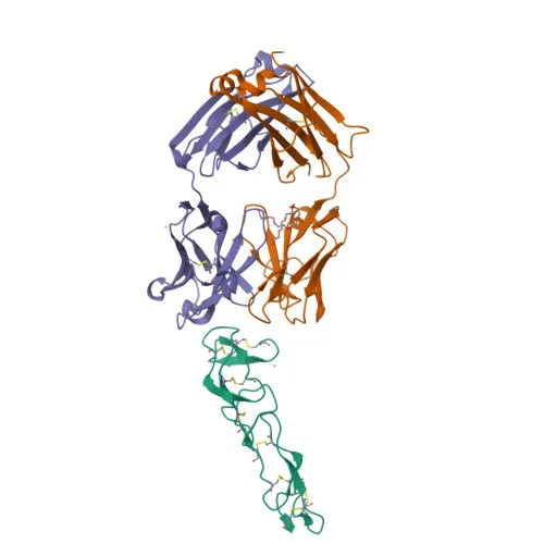
Monoclonal Antibodies
PerCP/Cyanine5.5 Rabbit anti-Human CD27 Monoclonal Antibody for Flow Cytometry - P26842
Item Number : CM0009253
Price varies based on specs and customizations
- Application
- FC
- Cross Reactivity
- Human
- Protein Weight
- 29kDa

Shipping:
Contact us to get shipping details Enjoy On-time Dispatch Guarantee.
Why Choose Us
Easy ordering process, quality products, and dedicated support for your business success.
Core Product Specifications and Parameters
| Parameter | Value |
|---|---|
| Product Name | PerCP/Cyanine5.5 Rabbit anti-Human CD27 mAb |
| Remarks/Alias | T14; S152; Tp55; TNFRSF7; S152. LPFS2 |
| Species | Human |
| Gene ID (Human) | 939 |
| Gene ID | 939 |
| Immunogen | Recombinant fusion protein containing a sequence corresponding to amino acids 21-192 of human CD27 (NP_001233.2). |
| Source | Rabbit |
| Category | Monoclonal Antibodies |
| Application | FC |
| Cross Reactivity | Human |
| SWISS | P26842 |
| Protein Weight | 29kDa |
| Shipping | Ice bag |
Biological Background: CD27 Function and Localization
- CD27 is a costimulatory immune-checkpoint receptor of the tumor necrosis factor receptor superfamily, expressed on the surface of T-cells, NK-cells, and B-cells. Related references: PMID:28011863
- It binds to its ligand CD70 (CD27L) expressed by B-cells, mediating antigen-specific T-cell activation and expansion, thereby providing immune surveillance of B-cells. Related references: PMID:28011863
- Upon CD70 ligation, CD27 activates the TRAF2-PTPN6 axis to inhibit LCK phosphorylation, driving T-cell memory adaptations. Related references: PMID:38354704
- CD27 provides a negative feedback signal to leukocyte differentiation during immune activation, thus modulating hematopoiesis.
- In adipose tissue, CD27 negatively regulates the function of Th2 lymphocytes.
- CD27 is localized at the cell membrane and is found in most T-lymphocytes.
- The protein undergoes phosphorylation and glycosylation, contains disulfide bonds, and features a transmembrane helix; it is associated with apoptosis signaling.
Experimental Guidance and Technical Tips
- The immunogen corresponds to the extracellular domain (aa 21–192), making this antibody suitable for detecting surface CD27 by flow cytometry.
- Consider titrating the antibody in your flow cytometry protocol to determine the optimal concentration for your cell type and experimental conditions.
- Include appropriate compensation controls when using PerCP/Cyanine5.5 to ensure accurate multicolor flow cytometry data.
- Validate the antibody in your specific sample system, as staining patterns may vary with activation state or cell lineage.
CamelBio: Your One-Stop Sourcing Bridge
This anti-CD27 monoclonal antibody supports flow cytometric analysis of T-cell subsets and immune monitoring applications. CamelBio provides diagnostic manufacturers, labs, and research institutes with one-stop access to IVD raw materials, including validated antibody pairs, optimized monoclonal/polyclonal antibodies, and bulk ancillary reagents. For CD27-related immuno-oncology research, we offer targeted sourcing solutions to meet your development needs from concept to clinic.
Product Datasheet
PerCP/Cyanine5.5 Rabbit anti-Human CD27 Monoclonal Antibody for Flow Cytometry - P26842
REQUEST A QUOTE
Our professional team will reply to you within one business day. Please feel free to contact us!