Monoclonal Antibodies
PE/Cyanine7 Rabbit anti-Mouse CD48 Monoclonal Antibody (Flow Cytometry) - P18181
Item Number : CM0008031
Price varies based on specs and customizations
- Application
- FC
- Cross Reactivity
- Mouse
- Protein Weight
- 27kDa

Shipping:
Contact us to get shipping details Enjoy On-time Dispatch Guarantee.
Why Choose Us
Easy ordering process, quality products, and dedicated support for your business success.
Core Product Specifications and Parameters
| Parameter | Specification |
|---|---|
| Product Name | PE/Cyanine7 Rabbit anti-Mouse CD48 mAb |
| Remarks/Alias | BCM1; BLAST; Bcm-1; BLAST1; SLAMF2; Sgp-60; BLAST-1; MEM-102 |
| Species | Mouse |
| Gene ID (Human) | 962 |
| Gene ID | 12506 |
| Immunogen | Recombinant protein|Recombinant fusion protein containing a sequence corresponding to amino acids 23-216 of mouse CD48 (NP_031675.1). |
| Source | Rabbit |
| Category | Monoclonal Antibodies |
| Application | FC |
| Cross Reactivity | Mouse |
| SWISS | P18181 |
| Protein Weight | 27kDa |
| Shipping | Ice bag |
Biological Background: CD48 Function and Localization
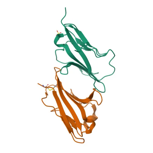
- CD48 (also known as BCM1, BLAST-1, SLAMF2, sgp-60) is a glycosylphosphatidylinositol (GPI)-anchored cell surface glycoprotein belonging to the signaling lymphocytic activation molecule (SLAM) family.
- It mediates immune cell activation and regulation by interacting with cell surface receptors such as 2B4/CD244 and CD2 via its N-terminal immunoglobulin domain. Related references: PMID:21278219, PMID:9881969
- In T-cells, CD48 associates with CD2 to recruit the Src kinase LCK and the adaptor LAT to the TCR/CD3 complex, enhancing LCK phosphorylation and downstream signaling. Related references: PMID:9881969
- Engagement of CD244 by CD48 induces phosphorylation of ITSMs in CD244, leading to immunological synapse formation and directed secretion of lytic granules (perforin, granzymes) by cytotoxic T-lymphocytes and NK cells. Related references: PMID:21278219, PMID:9881969
- Subcellularly, CD48 is found on the cell membrane, enriched in membrane rafts, and can also be secreted in a soluble form.
- UniProt keywords include GPI-anchor, immunoglobulin domain, disulfide bond, glycoprotein, and lipoprotein, reflecting its structural features and membrane association.
Experimental Guidance and Technical Tips
- The immunogen (amino acids 23-216 of mouse CD48) corresponds to the predicted extracellular domain, so the antibody is likely to recognize the native surface antigen. For flow cytometry, titrate the antibody to determine the optimal staining concentration using a relevant cell line or primary cells (e.g., mouse splenocytes, thymocytes).
- Since CD48 is a GPI-anchored protein, live-cell surface staining is typically performed without permeabilization. Include appropriate isotype control and fluorescence-minus-one (FMO) controls to confirm specificity.
- PE/Cyanine7 is a tandem dye; protect from prolonged light exposure to prevent photobleaching. Fixation may alter tandem dye fluorescence, so verify compatibility if intracellular staining is required.
- CD48 is expressed on all hematopoietic cells except erythrocytes; use known positive (e.g., EL4, YAC-1) and negative controls to validate staining patterns.
CamelBio: Your One-Stop Sourcing Bridge
CamelBio offers comprehensive one-stop sourcing solutions for IVD raw materials, including validated research-grade monoclonal antibodies, recombinant proteins, and ancillary reagents. For CD48-targeted studies, CamelBio can support your development workflow with optimized antibody pairs, bulk conjugates, and custom reagent sourcing, helping accelerate your diagnostic and research applications from concept to clinic.
Product Datasheet
PE/Cyanine7 Rabbit anti-Mouse CD48 Monoclonal Antibody (Flow Cytometry) - P18181
REQUEST A QUOTE
Our professional team will reply to you within one business day. Please feel free to contact us!