Monoclonal Antibodies
PE/Cyanine7 Rabbit anti-Mouse CD223/LAG-3 Monoclonal Antibody for Flow Cytometry - Q61790
Item Number : CM0004877
Price varies based on specs and customizations
- Application
- FC
- Cross Reactivity
- Mouse
- Protein Weight
- 57 kDa

Shipping:
Contact us to get shipping details Enjoy On-time Dispatch Guarantee.
Why Choose Us
Easy ordering process, quality products, and dedicated support for your business success.
Core Product Specifications and Parameters
| Parameter | Value |
|---|---|
| Product Name | PE/Cyanine7 Rabbit anti-Mouse CD223/LAG-3 mAb |
| Remarks/Alias | Ly66; CD223; LAG-3 |
| Species | Mouse |
| GeneID (Human) | 3902 |
| GeneID | 16768 |
| Immunogen | Recombinant fusion protein containing a sequence corresponding to amino acids 24-442 of mouse CD223/LAG-3 (NP_032505.1). |
| Source | Rabbit |
| Category | Monoclonal Antibodies |
| Application | FC |
| Cross Reactivity | Mouse |
| SWISS | Q61790 |
| Protein Weight | 57 kDa |
| Shipping | Ice bag |
Biological Background: CD223/LAG-3 Function and Localization
- LAG-3 acts as an inhibitory receptor on antigen-activated T cells, negatively regulating their proliferation, activation, effector function, and homeostasis upon binding to its main ligand MHC class II on antigen-presenting cells (APCs) and to FGL1 secreted by hepatocytes and certain tumor cells. Related references: PMID:12209638, PMID:12421911, PMID:12672063, PMID:30349037
- Mechanistically, ligand engagement promotes ubiquitination of the KIEELE motif, releasing the RRFSALE motif from the membrane and facilitating the formation of condensates with CD3E, which disrupts CD3E-LCK association and inhibits TCR signaling. Related references: PMID:40101708, PMID:40592325
- LAG-3 may synergize with PD-1 to inhibit antigen-specific T-cell activation. Related references: PMID:21300912
- LAG-3 contributes to immune tolerance by being constitutively expressed on a subset of regulatory T cells (Tregs), supporting their suppressive function, and also acts as a negative regulator of plasmacytoid dendritic cell (pDC) activation. Related references: PMID:15485628, PMID:19201841
- The LAG-3 inhibitory pathway is exploited by tumors to evade anti-tumor immunity.
- Additionally, LAG-3 may function as a ligand for MHC class II on APCs, promoting APC activation and driving Th1 immune responses.
- LAG-3 is primarily expressed on activated CD4+ and CD8+ T cells, as well as on Tregs and pDCs, and localizes at the cell membrane and is also secreted.
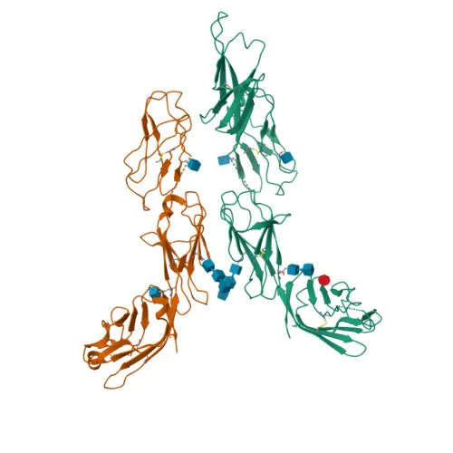
- Key structural features include immunoglobulin domains, disulfide bonds, and N-linked glycosylation, consistent with UniProt keywords: 3D-structure, Adaptive immunity, Cell membrane, Disulfide bond, Glycoprotein, Immunity, Immunoglobulin domain, Isopeptide bond, Membrane, Reference proteome, Repeat, Secreted, Signal, Transmembrane, Transmembrane helix, Ubl conjugation.
Experimental Guidance and Technical Tips
- The immunogen corresponds to the extracellular domain (aa 24-442) of mouse LAG-3, suggesting the antibody is suitable for detecting surface LAG-3 in live cell flow cytometry. Consider appropriate isotype controls and Fc receptor blocking to minimize background.
- Optimize antibody titration for your specific cell type and experimental conditions to achieve the best signal-to-noise ratio.
- Cross-reactivity has been confirmed for mouse; if using other species, validate specificity in your system.
CamelBio: Your One-Stop Sourcing Bridge
CamelBio provides diagnostic manufacturers and research institutions with a comprehensive one-stop platform for IVD raw materials, from validated antibody pairs and optimized monoclonal/polyclonal antibodies to bulk ancillary reagents. This anti-mouse LAG-3 antibody supports immune checkpoint research and T-cell exhaustion studies, and CamelBio can also help source rare-target reagents and custom services to accelerate your assay development. Reach out to let us streamline your procurement from concept to clinic.
Product Datasheet
PE/Cyanine7 Rabbit anti-Mouse CD223/LAG-3 Monoclonal Antibody for Flow Cytometry - Q61790
REQUEST A QUOTE
Our professional team will reply to you within one business day. Please feel free to contact us!