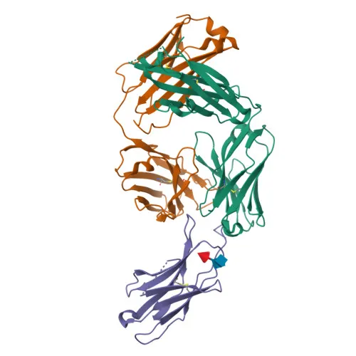
Monoclonal Antibodies
PE/Cyanine7 Rabbit anti-Human/Monkey PD-1/CD279 Monoclonal Antibody for FC - Q15116
Item Number : CM0012785
Price varies based on specs and customizations
- Application
- FC
- Cross Reactivity
- Human, Cynomolgus
- Protein Weight
- 32kDa

Shipping:
Contact us to get shipping details Enjoy On-time Dispatch Guarantee.
Why Choose Us
Easy ordering process, quality products, and dedicated support for your business success.
Core Product Specifications and Parameters
| Parameter | Value |
|---|---|
| Product Name | PE/Cyanine7 Rabbit anti-Human/Monkey PD-1/CD279 mAb |
| Remarks/Alias | PD1; PD-1; CD279; SLEB2; hPD-1; hPD-l; hSLE1 |
| Species | Human |
| GeneID (Human) | 5133 |
| GeneID | 5133 |
| Immunogen | Recombinant fusion protein containing a sequence corresponding to amino acids 25-168 of human PD-1/CD279 (NP_005009.2) |
| Source | Rabbit |
| Category | Monoclonal Antibodies |
| Application | FC |
| Cross Reactivity | Human, Cynomolgus |
| SWISS | Q15116 |
| Protein Weight | 32kDa |
| Shipping | Ice bag |
Biological Background: PD-1 Function and Localization
- PD-1 (Programmed cell death protein 1), also known as CD279, is an inhibitory receptor encoded by the PDCD1 gene that plays a critical role in induction and maintenance of immune tolerance. Related references: PMID:21276005, PMID:31754127, PMID:32184441
- It delivers inhibitory signals upon binding to its ligands CD274/PDCD1L1 and CD273/PDCD1LG2, downregulating T-cell responses. Related references: PMID:21276005, PMID:26602187
- Following T-cell receptor (TCR) engagement, PD-1 associates with TCR-CD3 in the immunological synapse and directly inhibits T-cell activation by recruiting PTPN11/SHP-2. Related references: PMID:32184441
- The SHP-2 phosphatase dephosphorylates key TCR proximal signaling molecules, including ZAP70, PRKCQ/PKCθ, and CD247/CD3ζ, thereby suppressing T-cell activation. Related references: PMID:32184441
- The PD-1-mediated inhibitory pathway is exploited by tumors to attenuate anti-tumor immunity; blockade of this pathway reverses exhausted T-cell phenotypes and restores anti-tumor responses, forming the basis of cancer immunotherapy. Related references: PMID:28951311, PMID:22658127, PMID:25034862, PMID:25399552
- PD-1 is a single-pass transmembrane glycoprotein localized to the cell membrane and contains an immunoglobulin-like domain, disulfide bonds, and intracellular signaling motifs subject to phosphorylation and ubiquitination.
Experimental Guidance and Technical Tips
- The immunogen used spans the extracellular domain (amino acids 25-168) of human PD-1, suggesting the antibody targets the native cell-surface conformation.
- When using this PE/Cyanine7-conjugated antibody for flow cytometry, ensure proper compensation settings and include appropriate isotype controls to account for non-specific binding.
- Cross-reactivity with cynomolgus monkey (Macaca fascicularis) PD-1 has been observed, potentially enabling translational studies in non-human primate models.
- It is advisable to titrate the antibody and validate its performance in the relevant sample system, as optimal conditions may vary depending on cell type and experimental setup.
CamelBio: Your One-Stop Sourcing Bridge
CamelBio offers diagnostic manufacturers, labs, and research institutes a streamlined one-stop channel for IVD raw materials, covering validated antibody pairs, optimized monoclonal and polyclonal antibodies, bulk ancillary reagents, and rare target sourcing. For PD-1/immune checkpoint research and immunotherapy development, CamelBio can supply this conjugated anti-PD-1 antibody alongside complementary reagents to support your discovery and translational workflows. Our integrated supply model helps you move from concept to clinic with reliable material access and expert consultation.
Product Datasheet
PE/Cyanine7 Rabbit anti-Human/Monkey PD-1/CD279 Monoclonal Antibody for FC - Q15116
REQUEST A QUOTE
Our professional team will reply to you within one business day. Please feel free to contact us!