Monoclonal Antibodies
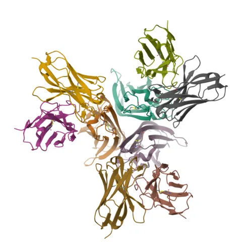
PE/Cyanine7 Rabbit anti-Human TNF-α Monoclonal Antibody for FC (intra) - P01375
Item Number : CM0005069
Price varies based on specs and customizations
- Application
- FC (intra)
- Cross Reactivity
- Human
- Protein Weight
- 26kDa

Shipping:
Contact us to get shipping details Enjoy On-time Dispatch Guarantee.
Why Choose Us
Easy ordering process, quality products, and dedicated support for your business success.
Core Product Specifications and Parameters
| Parameter | Value |
|---|---|
| Product Name | PE/Cyanine7 Rabbit anti-Human TNF-α mAb |
| Remarks/Alias | DIF; TNFA; TNFSF2; TNLG1F; TNF-alpha |
| Species | Human |
| GeneID (Human) | 7124 |
| GeneID | 7124 |
| Immunogen | Recombinant fusion protein containing a sequence corresponding to amino acids 77-233 of human TNF-α (NP_002344.2) |
| Source | Rabbit |
| Category | Monoclonal Antibodies |
| Application | FC (intra) |
| Cross Reactivity | Human |
| SWISS | P01375 |
| Protein Weight | 26kDa |
| Shipping | Ice bag |
Biological Background: TNF Function and Localization
- TNF-α (cachectin, TNF-alpha, TNFSF2) is a pleiotropic pro-inflammatory cytokine belonging to the tumor necrosis factor ligand superfamily, existing as both a type II transmembrane protein and a soluble homotrimer after proteolytic cleavage.
- It is primarily secreted by activated macrophages and binds to its receptors TNFRSF1A/TNFR1 and TNFRSF1B/TNFBR, triggering signaling pathways that can lead to cell death, survival, differentiation, or proliferation depending on the cellular context.
- TNF-α acts as an endogenous pyrogen, inducing fever directly or via IL-1 stimulation, and is a key mediator of cachexia in chronic inflammation and cancer.
- In regulatory T cells (Tregs), TNF-α impairs suppressive function by up-regulating protein phosphatase 1 (PP1), which dephosphorylates FOXP3 at Ser-418, thereby inactivating FOXP3 and compromising Treg activity in rheumatoid arthritis. Related references: PMID:23396208
- TNF-α can mediate anticancer activity: in bladder cancer cells, BCG-stimulated neutrophils kill tumor cells via a TNF-α-dependent mechanism that synergizes with SMAC mimetics. Related references: PMID:16829952 PMID:22517918 PMID:23396208
- It promotes angiogenesis by synergistically inducing VEGF production with IL-1β and IL-6, and it stimulates osteoclastogenesis, thereby contributing to bone resorption. Related references: PMID:12794819
- The intracellular domain (ICD) of TNF-α can be generated by regulated intramembrane proteolysis and has been shown to induce IL-12 production in dendritic cells.
- TNF-α is localized at the cell membrane and is also secreted; post-translational modifications include glycosylation, phosphorylation, myristoylation, and disulfide bond formation, which modulate its stability and function.
Experimental Guidance and Technical Tips
- The immunogen (aa 77-233) corresponds to the soluble extracellular domain of human TNF-α; this antibody is expected to recognize both the transmembrane precursor and the secreted mature cytokine.
- For intracellular flow cytometry (FC intra), optimize cell fixation and permeabilization conditions. Use unstimulated cells or irrelevant stimulations as negative controls.
- Titrate the antibody to determine the optimal staining concentration for your specific experimental system and cell type.
- Since cross-reactivity is limited to human, validate specificity in human samples and consider using species‑matched biological controls.
CamelBio: Your One-Stop Sourcing Bridge
As a comprehensive IVD raw-material partner, CamelBio connects diagnostic manufacturers, clinical laboratories, and research institutes with high-quality reagents like this TNF-α monoclonal antibody. We provide one-stop access to validated antibody pairs, optimized monoclonals and polyclonals, bulk ancillary reagents, and rare-target sourcing—supporting your immunology, inflammation, and oncology assay development from concept to clinic.
REQUEST A QUOTE
Our professional team will reply to you within one business day. Please feel free to contact us!