Monoclonal Antibodies
PE/Cyanine7 Rabbit anti-Human FcεR1α Monoclonal Antibody for FC - P12319
Item Number : CM0010229
Price varies based on specs and customizations
- Application
- FC
- Cross Reactivity
- Human
- Protein Weight
- 30kDa

Shipping:
Contact us to get shipping details Enjoy On-time Dispatch Guarantee.
Why Choose Us
Easy ordering process, quality products, and dedicated support for your business success.
Core Product Specifications and Parameters
| Parameter | Value |
|---|---|
| Product Name | PE/Cyanine7 Rabbit anti-Human FcεR1α mAb |
| Remarks/Alias | FCE1A; FcERI; FCERIA |
| Species | Human |
| GeneID (Human) | 2205 |
| GeneID | 2205 |
| Immunogen | Recombinant fusion protein containing a sequence corresponding to amino acids 26-205 of human FcεR1α (NP_001992.1). |
| Source | Rabbit |
| Category | Monoclonal Antibodies |
| Application | FC |
| Cross Reactivity | Human |
| SWISS | P12319 |
| Protein Weight | 30kDa |
| Shipping | Ice bag |
Biological Background: FcεR1α Function and Localization
- Protein identity: High affinity immunoglobulin epsilon receptor subunit alpha (FcεRIα), also known as Fc-epsilon RI-alpha or IgE Fc receptor subunit alpha, encoded by the FCER1A (FCE1A) gene.
- Core function: Serves as the high-affinity receptor for immunoglobulin E (IgE). Upon IgE binding and subsequent allergen cross-linking, it triggers signaling cascades leading to myeloid cell activation, degranulation, and release of vasoactive amines, lipid mediators, and cytokines.
- Immune response role: Mediates immediate hypersensitivity reactions to allergens and acts as a host defense mechanism against helminth parasites, pathogenic bacteria, and venom toxicity.
- Subcellular localization: Cell membrane; single-pass type I membrane protein.
- Tissue specificity: Expressed in eosinophils, mast cells, basophils, and other myeloid cells.
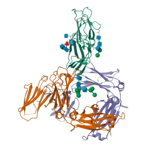
- Structural features: Glycoprotein with immunoglobulin-like domains, disulfide bonds, and a transmembrane helix. Belongs to the immunoglobulin superfamily.
- Disease relevance: Dysregulation of FcεRIα signaling can lead to severe allergic and anaphylactic reactions.
- Molecular weight: Approximately 30 kDa (glycosylated form) as observed by SDS-PAGE; the predicted unmodified mass is ~29.6 kDa.
Experimental Guidance and Technical Tips
- This antibody is directly conjugated to PE/Cyanine7 for flow cytometry (FC) applications, minimizing the need for secondary staining.
- The immunogen corresponds to the extracellular domain (amino acids 26–205) of human FcεR1α, ensuring recognition of the native cell surface receptor.
- Titrate the antibody to determine optimal staining concentration for your specific cell type, and include appropriate isotype controls.
- Validate specificity using FcεR1α-expressing cells such as eosinophils or basophils, or transfected cell lines.
- For multicolor FC panels, assess spectral overlap with other fluorochromes and design appropriate compensation controls.
CamelBio: Your One-Stop Sourcing Bridge
CamelBio provides comprehensive one-stop sourcing of IVD raw materials for diagnostic manufacturers, research laboratories, and institutes—covering every stage from concept to clinic. This anti-FcεR1α antibody supports allergy and inflammation research, backed by our reliable supply chains for monoclonal/polyclonal antibodies, bulk ancillary reagents, and rare target raw-material procurement. Contact us for validated antibody pairs, optimized conjugation services, and tailored sourcing solutions to accelerate your assay development.
REQUEST A QUOTE
Our professional team will reply to you within one business day. Please feel free to contact us!