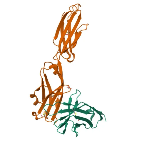
Monoclonal Antibodies
PE Rabbit anti-Mouse PD-L2/CD273/B7-DC Monoclonal Antibody for Flow Cytometry - Q9WUL5
Item Number : CM0009764
Price varies based on specs and customizations
- Application
- FC
- Cross Reactivity
- Mouse
- Protein Weight
- 28kDa

Shipping:
Contact us to get shipping details Enjoy On-time Dispatch Guarantee.
Why Choose Us
Easy ordering process, quality products, and dedicated support for your business success.
Core Product Specifications and Parameters
| Parameter | Specification |
|---|---|
| Product Name | PE Rabbit anti-Mouse PD-L2/CD273/B7-DC mAb |
| Remarks/Alias | Btdc; B7-DC; PD-L2; F730015O22Rik |
| Species | Mouse |
| Gene ID (Human) | 80380 |
| Gene ID | 58205 |
| Immunogen | Recombinant fusion protein containing a sequence corresponding to amino acids 20-123 of mouse PD-L2/CD273/B7-DC (NP_067371.1). |
| Source | Rabbit |
| Category | Monoclonal Antibodies |
| Application | FC |
| Cross Reactivity | Mouse |
| SWISS | Q9WUL5 |
| Protein Weight | 28kDa |
| Shipping | Ice bag |
Biological Background: PD-L2/CD273/B7-DC Function and Localization
- PD-L2 (Programmed cell death 1 ligand 2), also referred to as butyrophilin B7-DC (B7-DC) or CD273, is a transmembrane glycoprotein of the immunoglobulin superfamily, containing immunoglobulin-like domains and disulfide bonds.
- It plays a critical role in the induction and maintenance of immune tolerance to self, acting as a ligand for the inhibitory receptor PD-1 (PDCD1) on T cells, thereby inhibiting T-cell proliferation by blocking cell cycle progression and cytokine production. Related references: PMID:11224527 PMID:11283156 PMID:12719480
- The protein is localized to the cell membrane and possesses a single transmembrane helix (UniProt keyword).
- Tissue expression studies in mice show high levels in placenta and liver, with weaker expression in heart, spleen, lymph nodes, and thymus. It is also expressed in immature and mature bone marrow-derived dendritic cells and splenic dendritic cells, and some lymphoid-origin tumor cell lines.
- Post-translational modifications include glycosylation, and the protein contains a signal peptide, an immunoglobulin domain, and intramolecular disulfide bonds, critical for structural integrity and receptor binding.
- Keywords also indicate involvement in adaptive immunity and receptor activity, highlighting its role in immune checkpoint regulation.
Experimental Guidance and Technical Tips
- The immunogen consists of a recombinant fusion protein covering the extracellular domain (amino acids 20-123) of mouse PD-L2, which is expected to generate an antibody recognizing the native protein in flow cytometry applications.
- For flow cytometry, consider titrating the antibody to determine optimal staining concentration in the relevant cell type, such as mouse dendritic cells or PD-L2-transfected cell lines.
- Include appropriate isotype controls and viability dyes to ensure specificity and accurate gating.
- Validate the reactivity in your sample system, as cross-reactivity is only confirmed for mouse.
CamelBio: Your One-Stop Sourcing Bridge
As a comprehensive IVD raw-material sourcing platform, CamelBio connects diagnostic manufacturers and researchers with high-quality reagents like this anti-PD-L2 monoclonal antibody. We offer validated antibody pairs, optimized monoclonal/polyclonal antibodies, and bulk ancillary reagents to streamline your immunoassay development. Partner with us for efficient procurement and expert support in your immunology and immune checkpoint research projects.
Product Datasheet
PE Rabbit anti-Mouse PD-L2/CD273/B7-DC Monoclonal Antibody for Flow Cytometry - Q9WUL5
REQUEST A QUOTE
Our professional team will reply to you within one business day. Please feel free to contact us!