Monoclonal Antibodies
PE Rabbit anti-Mouse CD80 mAb for FC - Q00609
Item Number : CM0009522
Price varies based on specs and customizations
- Application
- FC
- Cross Reactivity
- Mouse
- Protein Weight
- 34kDa

Shipping:
Contact us to get shipping details Enjoy On-time Dispatch Guarantee.
Why Choose Us
Easy ordering process, quality products, and dedicated support for your business success.
Core Product Specifications and Parameters
| Parameter | Value |
|---|---|
| Product Name | PE Rabbit anti-Mouse CD80 mAb |
| Remarks/Alias | B71; Ly53; TSA1; Cd28l; Ly-53; MIC17 |
| Species | Mouse |
| GeneID (Human) | 941 |
| GeneID | 12519 |
| Immunogen | Recombinant protein (Recombinant fusion protein containing a sequence corresponding to amino acids 38-245 of mouse CD80, NP_033985.3) |
| Source | Rabbit |
| Category | Monoclonal Antibodies |
| Application | FC |
| Cross Reactivity | Mouse |
| SWISS | Q00609 |
| Protein Weight | 34kDa |
| Shipping | Ice bag |
Biological Background: CD80 Function and Localization
- CD80, also known as B7-1, is a member of the immunoglobulin superfamily. It functions as a costimulatory molecule essential for T-lymphocyte activation, providing a critical secondary signal alongside MHC/TCR engagement. Related references: PMID:16339518 PMID:1714935
- CD80 serves as a ligand for CD28, constitutively expressed on naive T-cells, triggering pathways like NF-kappa-B and MAPK to promote cytokine production. It also binds CTLA4, acting as an inhibitor to limit T-cell responses.
- Engagement of CD28 by CD80 stimulates glucose metabolism and ATP synthesis in T-cells via the PI3K/Akt signaling pathway, enhancing cellular energy for immune function.
- CD80 regulates PDL1/PDCD1 interactions, preventing excessive inhibitory signaling and balancing immune responses.
- On B-cells, CD80 is crucial for germinal center reactions, facilitating productive B-cell–T-cell interactions necessary for humoral immunity. Related references: PMID:22450810
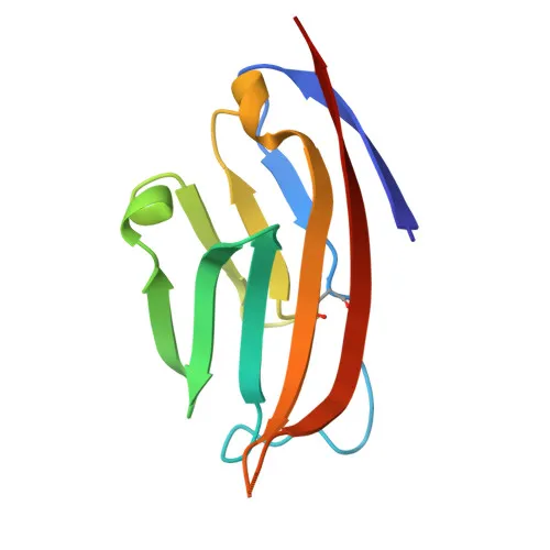
- CD80 is a single-pass type I membrane protein localized to the cell membrane. It contains an extracellular region with immunoglobulin-like domains, a transmembrane helix, and a short cytoplasmic tail.
- Post-translational modifications include N-linked glycosylation and disulfide bond formation, which are important for its structure and ligand interactions. The gene undergoes alternative splicing.
- Tissue expression is primarily on activated B-cells, gamma interferon-stimulated monocytes, and some non-circulating B-cell malignancies.
Experimental Guidance and Technical Tips
- The immunogen corresponds to the extracellular domain (aa 38–245) of mouse CD80, designed to target the native conformation accessible on the cell surface. Consider using this antibody in flow cytometry for detecting CD80 on activated immune cells.
- Since the antibody is PE-conjugated, optimize the staining protocol to avoid over-incubation and use appropriate isotype controls to minimize non-specific binding. Validate in the relevant mouse sample system, such as splenocytes or lymph node cells, under activation conditions.
- Given the role of CD80 in T-cell/B-cell interactions, this antibody may be useful in studying immune synapse formation or costimulatory blockade, but empirical validation is recommended.
CamelBio: Your One-Stop Sourcing Bridge
CamelBio streamlines the procurement of high-quality IVD raw materials for diagnostic manufacturers and research laboratories. This PE Rabbit anti-Mouse CD80 mAb supports immunology and immuno-oncology assay development, and we offer a comprehensive portfolio of validated antibody pairs, optimized monoclonal antibodies, and ancillary reagents. Leverage our supply chain to efficiently source rare targets or bulk quantities, accelerating your project from concept to clinic.
REQUEST A QUOTE
Our professional team will reply to you within one business day. Please feel free to contact us!