Monoclonal Antibodies
PE Rabbit anti-Mouse CD366 (TIM-3) Monoclonal Antibody for Flow Cytometry - Q8VIM0
Item Number : CM0012169
Price varies based on specs and customizations
- Application
- FC
- Cross Reactivity
- Mouse
- Protein Weight
- 22kDa/31kDa

Shipping:
Contact us to get shipping details Enjoy On-time Dispatch Guarantee.
Why Choose Us
Easy ordering process, quality products, and dedicated support for your business success.
Core Product Specifications and Parameters
| Parameter | Value |
|---|---|
| Product Name | PE Rabbit anti-Mouse CD366 (TIM-3) mAb |
| Remarks/Alias | Tim3; TIM-3; Timd3 |
| Species | Mouse |
| GeneID (Human) | 84868 |
| GeneID | 171285 |
| Immunogen | Recombinant protein|Recombinant fusion protein containing a sequence corresponding to amino acids 20-191 of mouse CD366 (TIM-3) (NP_599011.2) |
| Source | Rabbit |
| Category | Monoclonal Antibodies |
| Application | FC |
| Cross Reactivity | Mouse |
| SWISS | Q8VIM0 |
| Protein Weight | 22kDa/31kDa |
| Shipping | Ice bag |
Biological Background: CD366/TIM-3 Function and Localization
- Encoded by the Havcr2 gene, also known as TIMD-3 or TIM-3. Alternative splicing reported. Belongs to the immunoglobulin superfamily.
- Functions as a cell surface receptor with context-dependent roles: generally inhibitory but can activate immune responses. Inhibitory functions include regulation of macrophage activation, promotion of immunological tolerance by suppressing Th1-mediated auto- and alloimmune responses, attenuation of TCR signaling in CD8+ T cells, and negative regulation of NK cell function during endotoxic shock. Related references: PMID:11823861 PMID:14556006 PMID:18006747 PMID:25337993
- Activating roles include enhancing TCR signaling in T-cells via ZAP70, LCP2, LCK and FYN; promoting antibacterial activity of Th1 cells against Mycobacterium tuberculosis; enhancing CD8+ T-cell responses to Listeria monocytogenes infection; and stimulating mast cell production of Th2 cytokines IL-4, IL-6 and IL-13. Also reported to act as an activating coreceptor on NK cells for IFN-gamma production (By similarity). Related references: PMID:21807895 PMID:20937702 PMID:24567532 PMID:17620455
- Serves as a receptor for galectin-9 (LGALS9): binding can induce T-cell apoptosis through HAVCR2 phosphorylation and disruption of BAG6 interaction. Also involved in innate immune response to intracellular pathogens via LGALS9 interaction on infected macrophages. However, the LGALS9 receptor function has been challenged (By similarity). Related references: PMID:22863785 PMID:20937702
- Functions as a phosphatidylserine (PtSer) receptor in a calcium-dependent manner. Mediates engulfment of apoptotic cells by dendritic cells and promotes T-cell conjugation to apoptotic cells without engulfment. Related references: PMID:20083673 PMID:19224762
- On dendritic cells, positively regulates innate immunity and synergizes with Toll-like receptors to promote TNF secretion. In tumor-infiltrating dendritic cells, suppresses nucleic acid-mediated innate immune responses by interacting with HMGB1 and interfering with nucleic acid sensing. Related references: PMID:18006747 PMID:22842346
- Localizes to the membrane and cell junctions, and a secreted form exists. Tissue expression: up-regulated on differentiated Th1 and CD8+ cytotoxic T cells, also on macrophages, monocytes, dendritic cells, NK cells, and mast cells; absent from naive T cells.
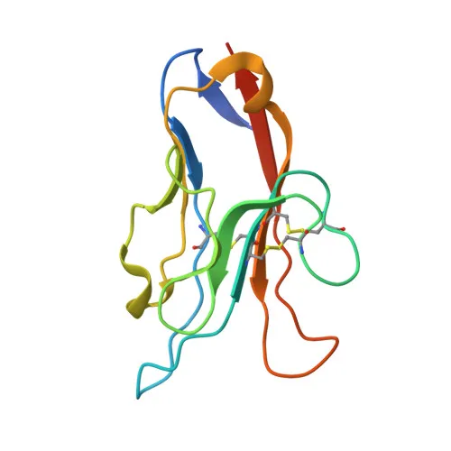
- Post-translational modifications: glycoprotein, disulfide bonds, phosphorylation. Contains an immunoglobulin domain.
Experimental Guidance and Technical Tips
- The immunogen corresponds to the extracellular domain of mouse TIM-3 (amino acids 20–191), which is suitable for flow cytometry applications targeting cell surface expression.
- Validate antibody performance in relevant mouse immune cell subsets (e.g., Th1, CD8+ T cells, macrophages, dendritic cells) where TIM-3 expression is reported under activating or tolerogenic conditions.
- PE conjugation enables direct detection without secondary antibody; ensure minimal light exposure and use appropriate compensation controls in multicolor flow cytometry panels. Consider titrating the antibody to determine the optimal staining index for your specific experimental setup.
CamelBio: Your One-Stop Sourcing Bridge
CamelBio supports diagnostic manufacturers, laboratories, and research institutes with comprehensive access to IVD raw materials, technical services, and consulting. Our portfolio includes validated monoclonal antibodies like this anti-TIM-3 clone, optimized for flow cytometry applications in immuno-oncology research. We also offer bulk ancillary reagents and facilitate sourcing of rare targets to accelerate your assay development from concept to clinic.
Product Datasheet
PE Rabbit anti-Mouse CD366 (TIM-3) Monoclonal Antibody for Flow Cytometry - Q8VIM0
REQUEST A QUOTE
Our professional team will reply to you within one business day. Please feel free to contact us!