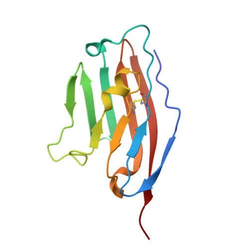
Monoclonal Antibodies
PE Rabbit anti-Mouse CD274/PD-L1 Monoclonal Antibody for Flow Cytometry - Q9EP73
Item Number : CM0011843
Price varies based on specs and customizations
- Application
- FC
- Cross Reactivity
- Mouse
- Protein Weight
- 32kDa

Shipping:
Contact us to get shipping details Enjoy On-time Dispatch Guarantee.
Why Choose Us
Easy ordering process, quality products, and dedicated support for your business success.
Core Product Specifications and Parameters
| Parameter | Value |
|---|---|
| Product Name | PE Rabbit anti-Mouse CD274/PD-L1 mAb |
| Remarks/Alias | B7h1, Pdl1, Pdcd1l1, Pdcd1lg1, A530045L16Rik |
| Species | Mouse |
| GeneID (Human) | 29126 |
| GeneID | 60533 |
| Immunogen | Recombinant protein |
| Source | Rabbit |
| Category | Monoclonal Antibodies |
| Application | FC |
| Cross Reactivity | Mouse |
| SWISS | Q9EP73 |
| Protein Weight | 32kDa |
| Shipping | Ice bag |
Biological Background: PD-L1 (CD274) Function and Localization
- PD-L1 plays a critical role in induction and maintenance of immune tolerance to self by binding to the inhibitory receptor PD-1 (PDCD1), thereby modulating T-cell activation thresholds and limiting T-cell effector responses. Related references: PMID:11238124
- Through a yet unknown activating receptor, PD-L1 may costimulate T-cell subsets that predominantly produce interleukin-10 (IL-10). Related references: PMID:11015443 PMID:12719480
- The PD-1/PD-L1 pathway is exploited by tumors to attenuate anti-tumor immunity; PD-L1 interaction with PD-1 inhibits cytotoxic T lymphocyte (CTL) effector function, and blocking this pathway can reverse T-cell exhaustion and normalize anti-tumor responses, providing a rationale for cancer immunotherapy. Related references: PMID:12218188 PMID:27281199
- PD-L1 (CD274), also known as B7 homolog 1 (B7-H1), is a member of the immunoglobulin superfamily and contains immunoglobulin-like domains.
- PD-L1 is localized to the cell membrane, early endosome membrane, and recycling endosome membrane.
- Tissue expression: Highly expressed in the heart, thymus, skeletal muscle, and lung; weakly expressed in the kidney, spleen, thyroid, and liver. It is expressed on activated dendritic cells, B-cells, and macrophages, and in numerous tumor cell lines of lymphoid origin.
- Post-translational modifications include glycosylation and disulfide bond formation, as indicated by UniProt keywords (glycoprotein, disulfide bond, Ubl conjugation).
Experimental Guidance and Technical Tips
- This PE-conjugated monoclonal antibody is designed for flow cytometry and likely recognizes the native conformation of mouse PD-L1 on the cell surface, given that the immunogen corresponds to the extracellular domain (amino acids 19-235).
- For flow cytometry, consider using appropriate isotype control antibodies and Fc receptor blocking to minimize nonspecific binding. Titrate the antibody for optimal signal-to-noise ratio in your experimental system.
- As a PE conjugate, ensure proper compensation controls when combined with other fluorophores in multicolor panels.
- Validate staining with relevant mouse cell lines or primary cells known to express PD-L1 (e.g., activated immune cells or tumor cell lines).
CamelBio: Your One-Stop Sourcing Bridge
CamelBio serves as your one-stop sourcing partner for IVD raw materials, supporting diagnostic manufacturers and researchers from concept to clinic. For PD-L1 and immuno-oncology research, we offer streamlined access to validated antibody pairs, optimized monoclonal/polyclonal antibodies, bulk ancillary reagents, and rare target raw-material sourcing. Our integrated supply capabilities help accelerate your assay development and diagnostic commercialization.
Product Datasheet
PE Rabbit anti-Mouse CD274/PD-L1 Monoclonal Antibody for Flow Cytometry - Q9EP73
REQUEST A QUOTE
Our professional team will reply to you within one business day. Please feel free to contact us!