Monoclonal Antibodies
PE Rabbit anti-Mouse CD1d Monoclonal Antibody for Flow Cytometry - P11609 / P11610
Item Number : CM0010023
Price varies based on specs and customizations
- Application
- FC
- Cross Reactivity
- Mouse
- Protein Weight
- 39kDa/38kDa

Shipping:
Contact us to get shipping details Enjoy On-time Dispatch Guarantee.
Why Choose Us
Easy ordering process, quality products, and dedicated support for your business success.
Core Product Specifications and Parameters
| Parameter | Value |
|---|---|
| Product Name | PE Rabbit anti-Mouse CD1d mAb |
| Remarks/Alias | Cd1a; Cd1d; CD1.1; Ly-38 |
| Species | Mouse |
| GeneID (Human) | 912 |
| GeneID (Mouse) | 12479/12480 |
| Immunogen | Recombinant fusion protein containing a sequence corresponding to amino acids 22-305 of mouse CD1d (NP_031665.2/NP_031666.2) |
| Source | Rabbit |
| Category | Monoclonal Antibodies |
| Application | FC |
| Cross Reactivity | Mouse |
| SWISS | P11609/P11610 |
| Protein Weight | 39kDa/38kDa |
| Shipping | Ice bag |
Biological Background: CD1d Function and Localization
- CD1d (cluster of differentiation 1d) is a member of the CD1 family of antigen-presenting molecules, encompassing two functional proteins in mice: CD1d1 (encoded by Cd1d1, SWISS: P11609) and CD1d2 (encoded by Cd1d2, SWISS: P11610).
- CD1d proteins are specialized in presenting self- and microbial-derived glycolipids to a subset of T cells known as natural killer T (NKT) cells, bridging innate and adaptive immunity.
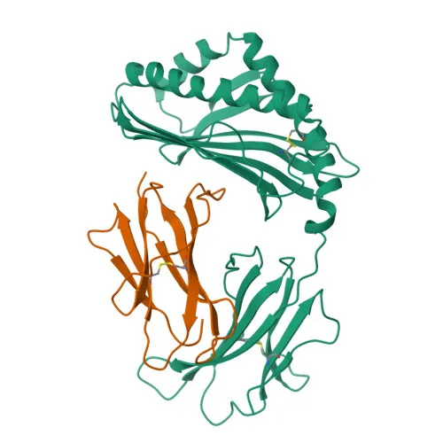
- CD1d is a type I transmembrane glycoprotein with an extracellular region containing an immunoglobulin-like domain, enabling antigen binding and presentation.
- Subcellularly, CD1d traffics between the cell surface and endosomal/lysosomal compartments, where it can sample and bind lipid antigens before returning to the plasma membrane for T-cell engagement.
- In mice, CD1d is expressed on the surface of cortical thymocytes, certain T-cell leukemias, and various other tissues, consistent with a role in immune surveillance.
- This rabbit monoclonal antibody recognizes both mouse CD1d isoforms and is suitable for studying NKT cell biology, lipid-based immunity, and related immunological pathways.
Experimental Guidance and Technical Tips
- The PE conjugate allows direct detection by flow cytometry, eliminating the need for secondary antibody incubation; optimal antibody titration should be determined empirically for each experimental system.
- Use appropriate isotype controls and include an Fc receptor blocking step to minimize non-specific binding in mouse samples.
- For surface staining, prepare single-cell suspensions from lymphoid organs (spleen, thymus, lymph nodes) or cultured cells and incubate with the antibody in staining buffer at recommended temperatures (typically 4°C for 30 minutes).
- CD1d may be expressed at relatively low levels on some cell types; using a bright fluorophore like PE can improve signal resolution.
CamelBio: Your One-Stop Sourcing Bridge
CamelBio provides diagnostic manufacturers, research laboratories, and academic institutions with reliable access to high-quality IVD raw materials, including monoclonal antibodies, recombinant proteins, and ancillary reagents. As an authorized channel for this PE-conjugated anti-CD1d antibody, we offer streamlined sourcing for immunology research focusing on NKT cells, lipid antigen presentation, and rare‑target antibody development. Whether you need validated antibody pairs for assay development or bulk custom formulations, CamelBio serves as your bridging partner from concept to clinical application.
Product Datasheet
PE Rabbit anti-Mouse CD1d Monoclonal Antibody for Flow Cytometry - P11609 / P11610
REQUEST A QUOTE
Our professional team will reply to you within one business day. Please feel free to contact us!