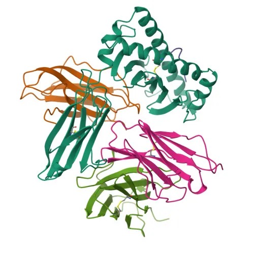
Monoclonal Antibodies
PE Rabbit anti-Human/Monkey CD8a Monoclonal Antibody for Flow Cytometry - P01732
Item Number : CM0004207
Price varies based on specs and customizations
- Application
- FC
- Cross Reactivity
- Human, Cynomolgus
- Protein Weight
- 21kDa/25kDa/30kDa

Shipping:
Contact us to get shipping details Enjoy On-time Dispatch Guarantee.
Why Choose Us
Easy ordering process, quality products, and dedicated support for your business success.
Core Product Specifications and Parameters
| Parameter | Value |
|---|---|
| Product Name | PE Rabbit anti-Human/Monkey CD8a mAb |
| Remarks/Alias | CD8; p32; Leu2; CD8alpha |
| Species | Human |
| Gene ID | 925 |
| Immunogen | Recombinant fusion protein containing a sequence corresponding to amino acids 22-182 of human CD8A (NP_001759.3) |
| Source | Rabbit |
| Category | Monoclonal Antibodies |
| Application | FC |
| Cross Reactivity | Human, Cynomolgus |
| SWISS | P01732 |
| Protein Weight | 21kDa/25kDa/30kDa |
| Shipping | Ice bag |
Biological Background: CD8a Function and Localization
- CD8a is an integral membrane glycoprotein that serves as a coreceptor for MHC class I molecules on T-cells, recognizing peptide antigens derived from cytosolic proteins.
- It interacts simultaneously with the T-cell receptor (TCR) and MHC class I, recruiting the Src kinase LCK to initiate signaling pathways leading to T-cell activation, proliferation, and cytotoxic responses.
- In natural killer (NK) cells, CD8A homodimers at the cell surface support conjugation and lysis of multiple target cells, and promote survival and differentiation into memory CD8 T-cells.
- The protein is predominantly expressed at the cell membrane but can also be secreted. Tissue expression includes thymus-derived T-cells (usually as a heterodimer with CD8B), NK cells, memory T-cells, intraepithelial lymphocytes, monocytes, and dendritic cells.
- It is also detected on plasmacytoid dendritic cells upon herpes simplex virus-1 stimulation.
- Post-translational modifications include glycosylation, disulfide bond formation, and palmitoylation, which are important for its stability and function.
- Alternative splicing produces multiple isoforms, and mutations are associated with disease susceptibility.
Experimental Guidance and Technical Tips
- The immunogen covers the extracellular domain (aa 22-182), making this antibody suitable for detecting native CD8a on live cells without permeabilization.
- For flow cytometry, titrate the antibody to determine the optimal concentration for your specific cell type and staining conditions, and include appropriate isotype controls.
- Validate antibody performance using known positive controls (e.g., human peripheral blood T-cells) and negative controls (e.g., CD8a-negative cell lines).
- Consider using this PE-conjugated antibody in multicolor panels with other lineage markers to characterize T-cell subsets.
CamelBio: Your One-Stop Sourcing Bridge
CamelBio provides diagnostic manufacturers and research laboratories with reliable access to IVD-critical raw materials, including validated monoclonal antibodies like this CD8a clone. Leverage our extensive supply network for bulk ancillary reagents, optimized antibody pairs, and rare-target sourcing—all designed to accelerate your immune-monitoring assay development. Let CamelBio be your procurement partner for CD8a-related research and beyond.
Product Datasheet
PE Rabbit anti-Human/Monkey CD8a Monoclonal Antibody for Flow Cytometry - P01732
REQUEST A QUOTE
Our professional team will reply to you within one business day. Please feel free to contact us!