Monoclonal Antibodies
PE Rabbit anti-Human TLR3 Monoclonal Antibody for Flow Cytometry (Intracellular) - O15455
Item Number : CM0008254
Price varies based on specs and customizations
- Application
- FC (intra)
- Cross Reactivity
- Human
- Protein Weight
- 104kDa

Shipping:
Contact us to get shipping details Enjoy On-time Dispatch Guarantee.
Why Choose Us
Easy ordering process, quality products, and dedicated support for your business success.
Core Product Specifications and Parameters
| Parameter | Value |
|---|---|
| Product Name | PE Rabbit anti-Human TLR3 mAb |
| Remarks/Alias | CD283; IIAE2; IMD83 |
| Species | Human |
| Gene ID (Human) | 7098 |
| Gene ID | 7098 |
| Immunogen | Recombinant protein corresponding to a sequence within amino acids 24-339 of human TLR3 (NP_003256.1) |
| Source | Rabbit |
| Category | Monoclonal Antibodies |
| Application | FC (intra) |
| Cross Reactivity | Human |
| SWISS | O15455 |
| Protein Weight | 104kDa |
| Shipping | Ice bag |
Biological Background: TLR3 Function and Localization
- Toll-like receptor 3 (TLR3), also known as CD283, is a single-pass type I membrane protein belonging to the Toll-like receptor family of pattern recognition receptors.
- Acts as a key sensor of viral double-stranded RNA (dsRNA), triggering innate immune responses via the TRIF/TICAM1 adaptor pathway, leading to NF-κB activation and IRF3 nuclear translocation, and subsequent pro-inflammatory cytokine and type I interferon production.
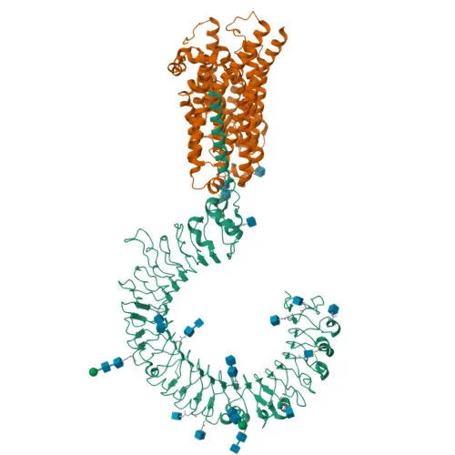
- The protein contains an extracellular leucine-rich repeat (LRR) domain responsible for ligand recognition, a transmembrane helix, and a cytoplasmic TIR (Toll/interleukin-1 receptor) domain essential for downstream signaling.
- Localizes primarily to the endoplasmic reticulum membrane and endosomal compartments (including early endosomes), where it encounters internalized viral dsRNA.
- TLR3 is highly expressed in the placenta and pancreas, and is the most strongly expressed TLR in the brain, notably in astrocytes, glia, and neurons. It is also found in CD11c+ immature dendritic cells.
- Post-translational modifications include N-linked glycosylation, phosphorylation, disulfide bond formation, and ubiquitination (isopeptide bond) — all contributing to receptor stability, trafficking, and signaling competence.
- Alternative splicing produces multiple isoforms, and genetic variants in TLR3 have been associated with disease susceptibility, including immune-related and neurological disorders.
Experimental Guidance and Technical Tips
- The antibody was generated against a recombinant fragment corresponding to the extracellular domain (aa 24-339). For intracellular flow cytometry, ensure proper cell fixation and permeabilization protocols to allow antibody access to the target protein.
- Consider titrating the antibody to determine the optimal concentration for your specific experimental setup, as staining efficiency may vary with cell type and fixation method.
- Use appropriate biological controls (e.g., unstimulated vs. poly(I:C)-stimulated cells) and isotype controls to verify staining specificity.
CamelBio: Your One-Stop Sourcing Bridge
CamelBio supports your innate immunity and infectious disease research with a comprehensive portfolio of IVD raw materials, including validated antibody pairs and optimized monoclonal antibodies like this PE-conjugated anti-TLR3 clone. From bulk ancillary reagents to rare target raw-material sourcing, we serve as your dedicated partner from concept to clinic.
Product Datasheet
PE Rabbit anti-Human TLR3 Monoclonal Antibody for Flow Cytometry (Intracellular) - O15455
REQUEST A QUOTE
Our professional team will reply to you within one business day. Please feel free to contact us!