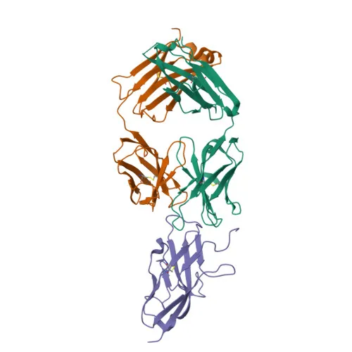
Monoclonal Antibodies
PE Rabbit anti-Human Siglec-8 Monoclonal Antibody for FC - Q9NYZ4
Item Number : CM0006216
Price varies based on specs and customizations
- Application
- FC
- Cross Reactivity
- Human
- Protein Weight
- 44kDa/46kDa/54kDa

Shipping:
Contact us to get shipping details Enjoy On-time Dispatch Guarantee.
Why Choose Us
Easy ordering process, quality products, and dedicated support for your business success.
Core Product Specifications and Parameters
| Parameter | Value |
|---|---|
| Product Name | PE Rabbit anti-Human Siglec-8 mAb |
| Remarks/Alias | SAF2; SIGLEC-8; SIGLEC8L |
| Species | Human |
| GeneID (Human) | 27181 |
| Immunogen | Recombinant fusion protein containing a sequence corresponding to amino acids 17-363 of human Siglec-8 (NP_055257.2) |
| Source | Rabbit |
| Category | Monoclonal Antibodies |
| Application | FC |
| Cross Reactivity | Human |
| SWISS | Q9NYZ4 |
| Protein Weight | 44kDa/46kDa/54kDa |
| Shipping | Ice bag |
Biological Background: Siglec-8 Function and Localization
- Siglec-8, also known as SAF-2 (sialoadhesin family member 2), is a single-pass type I membrane glycoprotein belonging to the immunoglobulin superfamily. Alternative splicing generates multiple isoforms.
- Siglec-8 mediates sialic-acid dependent adhesion to blood cells, preferentially binding α-2,3-linked sialic acid with some affinity for α-2,6-linkages; cis interactions on the same cell surface may mask the ligand-binding site. Related references: PMID:10625619, PMID:10856141
- It specifically recognizes terminal N-acetylneuraminic acid (sialic acid) and underlying 6-O-sulfated galactose, with a strong preference for Gal-6-sulfated sialyl-Lewis X glycan epitopes. Related references: PMID:27357658
- The protein is predominantly expressed on the surface of eosinophils, basophils, and mast cells, making it a specific marker for these cell types in hematopoietic studies.
- Subcellular localization is primarily at the plasma membrane, consistent with its role in cell-cell interactions and signaling.
- Siglec-8 contains immunoglobulin-like domains and disulfide bonds, typical of adhesion molecules, and is post-translationally modified by glycosylation.
Experimental Guidance and Technical Tips
- The immunogen corresponds to the extracellular domain (amino acids 17-363), suggesting the antibody likely recognizes surface-exposed epitopes, making it suitable for flow cytometry on live, non-permeabilized cells.
- For flow cytometry applications, titrate the PE-conjugated antibody to determine the optimal staining concentration using relevant cell lines or primary cells known to express Siglec-8 (e.g., eosinophil, basophil, or mast cell populations).
- Include appropriate isotype controls and Fc receptor blocking steps to minimize non-specific binding.
- Validate staining specificity in the intended sample system, considering the reported expression of Siglec-8 on human basophils, eosinophils, and mast cells.
- Note that multiple protein isoforms exist (44, 46, 54 kDa), but the PE conjugate is designed for cell surface detection and will not distinguish molecular weight variants.
CamelBio: Your One-Stop Sourcing Bridge
CamelBio provides comprehensive IVD raw-material solutions, offering validated antibody pairs, optimized monoclonal/polyclonal antibodies, and ancillary reagents to support your diagnostic and research workflows. This PE-conjugated anti-Siglec-8 antibody exemplifies our capability to deliver specialized immunoreagents for flow cytometric analysis, with a focus on rare and emerging targets in immunology and allergy research. Leverage our one-stop sourcing platform to secure high-quality raw materials and accelerate your development from concept to clinic.
REQUEST A QUOTE
Our professional team will reply to you within one business day. Please feel free to contact us!