Monoclonal Antibodies
PE Rabbit anti-Human MICA/MICB Monoclonal Antibody for FC - Q29983 / Q29980
Item Number : CM0005309
Price varies based on specs and customizations
- Application
- FC
- Cross Reactivity
- Human
- Protein Weight
- 4kDa/31kDa/37kDa/43kDa

Shipping:
Contact us to get shipping details Enjoy On-time Dispatch Guarantee.
Why Choose Us
Easy ordering process, quality products, and dedicated support for your business success.
Core Product Specifications and Parameters
| Parameter | Value |
|---|---|
| Product Name | PE Rabbit anti-Human MICA/MICB mAb |
| Remarks/Alias | MIC-A; PERB11.1; PERB11.2; MICB |
| Species | Human |
| GeneID (human) | 100507436/4277 |
| GeneID | 100507436/4277 |
| Immunogen | Recombinant protein | Recombinant fusion protein containing a sequence corresponding to amino acids 23-298 of human MICB (NP_005922.2). |
| Source | Rabbit |
| Category | Monoclonal Antibodies |
| Application | FC |
| Cross Reactivity | Human |
| SWISS | Q29983/Q29980 |
| Protein Weight | 4kDa/31kDa/37kDa/43kDa |
| Shipping | Ice bag |
Biological Background: MICA/MICB Function and Localization
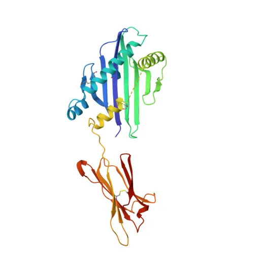
- MICA is a membrane-bound glycoprotein of the MHC class I-related family, containing immunoglobulin-like domains and structural features such as disulfide bonds and palmitoylation sites.
- MICA acts as a high-affinity ligand for the activating receptor NKG2D (KLRK1) on NK cells, γδ T cells, and CD8+ αβ T cells, mediating immune surveillance and antitumor responses. Related references: PMID:11491531, PMID:11777960
- Under cellular stress (e.g., infection, DNA damage), MICA expression is markedly upregulated, functioning as a stress signal that triggers NK cell cytotoxicity and cytokine production. Related references: PMID:10426993
- Subcellularly, MICA is primarily localized on the cell membrane but can also be detected in the cytoplasm.
- Tissue distribution is broad except in the central nervous system; highest expression is found in gastric epithelium, monocytes, keratinocytes, endothelial cells, and many epithelial tumors.
- Post-translational modifications of MICA include glycosylation, palmitoylation, and ubiquitination, which modulate its stability and immune recognition.
- MICA is subject to alternative splicing, generating multiple isoforms with potentially distinct functions.
- MICA interacts with viral proteins (host-virus interaction) and plays a role in viral immune evasion strategies.
Experimental Guidance and Technical Tips
- For flow cytometry, titrate the PE-conjugated antibody to determine the optimal concentration for your specific cell type and assay conditions.
- Use an appropriate isotype control antibody (e.g., PE-conjugated rabbit IgG) to assess non-specific binding.
- Protect the antibody and stained samples from light to prevent photobleaching of the PE fluorophore.
- Since the antibody recognizes both MICA and MICB, confirm target specificity in your experimental system by using cell lines with differential expression or through gene-specific knockdown.
- For cell surface staining, avoid fixation methods that may alter the epitope; if fixation is required, test compatibility with your protocol.
- MICA/MICB expression is elevated in stressed or transformed cells; include unstimulated or normal controls to establish basal expression levels.
CamelBio: Your One-Stop Sourcing Bridge
CamelBio serves as a comprehensive sourcing partner for IVD raw materials, offering this PE-conjugated anti-MICA/MICB monoclonal antibody to support immune-oncology and infectious disease research. With expertise in validated antibody pairs, bulk ancillary reagents, and rare target sourcing, we help diagnostic manufacturers and labs streamline assay development from concept to clinical application.
Product Datasheet
PE Rabbit anti-Human MICA/MICB Monoclonal Antibody for FC - Q29983 / Q29980
REQUEST A QUOTE
Our professional team will reply to you within one business day. Please feel free to contact us!