Monoclonal Antibodies
PE Rabbit anti-Human CD8 PolymAb® Monoclonal Antibody for FC - P01732 / P10966
Item Number : CM0010076
Price varies based on specs and customizations
- Application
- FC
- Cross Reactivity
- Human
- Protein Weight
- 26 kDa/24 kDa

Shipping:
Contact us to get shipping details Enjoy On-time Dispatch Guarantee.
Why Choose Us
Easy ordering process, quality products, and dedicated support for your business success.
Core Product Specifications and Parameters
| Parameter | Value |
|---|---|
| Product Name | PE Rabbit anti-Human CD8 PolymAb® |
| Remarks/Alias | CD8; p32; Leu2; IMD116; CD8alpha; LY3; P37; LEU2; LYT3; Ly-3; CD8B1; CD8beta. |
| Species | Human |
| GeneID | 925/926 |
| Source | Rabbit |
| Category | Monoclonal Antibodies |
| Application | FC |
| Cross Reactivity | Human |
| SWISS | P01732/P10966 |
| Protein Weight | 26 kDa/24 kDa |
| Shipping | Ice bag |
Biological Background: CD8 Function and Localization
- The CD8A gene encodes the T-cell surface glycoprotein CD8 alpha chain, also known as T-lymphocyte differentiation antigen T8/Leu-2.
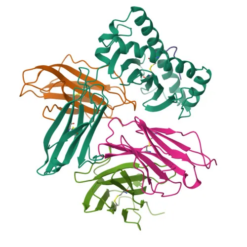
- CD8 functions as an integral membrane glycoprotein coreceptor for MHC class I molecules, binding simultaneously to the T-cell receptor (TCR) and MHC class I:peptide complex on antigen-presenting cells.
- Upon interaction with MHC class I, CD8 recruits the Src kinase LCK to the TCR-CD3 complex, initiating intracellular signaling that promotes lymphocyte activation, cytokine production, and cytotoxic T-cell (CTL) effector functions.
- CD8A is expressed on thymus-derived T-cells primarily as a disulfide-linked heterodimer with CD8B, but can also form CD8A homodimers on subsets of natural killer (NK) cells, memory T-cells, intraepithelial lymphocytes, monocytes, and dendritic cells.
- The CD8A homodimer on NK cells supports a survival mechanism that facilitates conjugation and lysis of multiple target cells, and also promotes differentiation of activated lymphocytes into memory CD8 T-cells.
- CD8A localizes to the cell membrane as a single-pass type I transmembrane protein; a secreted form has also been identified.
- Key post-translational modifications include alternative splicing, disulfide bond formation, N-linked glycosylation, and palmitoylation at a membrane-proximal cysteine residue, which are critical for its structure and function.
Experimental Guidance and Technical Tips
- For flow cytometric analysis, titrate the antibody to determine the optimal working dilution for your specific experimental system and cell type.
- Use appropriate isotype controls and fluorescence-minus-one (FMO) controls to assess background and set proper gating.
- Protect the PE-conjugated antibody from light during handling and storage to prevent photobleaching.
- This antibody detects both CD8A and CD8B chains; consider its suitability for identifying CD8+ T cell subsets and other CD8-expressing populations such as NK cells and dendritic cells in human peripheral blood mononuclear cells (PBMCs).
- Perform surface staining on live cells under standard conditions; avoid fixation before staining if possible, as epitope masking may occur.
CamelBio: Your One-Stop Sourcing Bridge
CamelBio is your one-stop partner for IVD raw materials, supporting immune research and diagnostic development. We supply validated anti-CD8 monoclonal antibodies, including PE conjugates, along with matched antibody pairs, ancillary reagents, and bulk raw materials for T-cell immunophenotyping and immune monitoring assays. Our sourcing capabilities extend to rare-target reagents and custom conjugations to accelerate your diagnostic pipeline.
Product Datasheet
PE Rabbit anti-Human CD8 PolymAb® Monoclonal Antibody for FC - P01732 / P10966
REQUEST A QUOTE
Our professional team will reply to you within one business day. Please feel free to contact us!