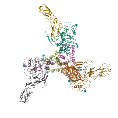
Monoclonal Antibodies
PE Rabbit anti-Human CD4 Monoclonal Antibody for FC - P01730
Item Number : CM0009094
Price varies based on specs and customizations
- Application
- FC
- Cross Reactivity
- Human
- Protein Weight
- 51kDa

Shipping:
Contact us to get shipping details Enjoy On-time Dispatch Guarantee.
Why Choose Us
Easy ordering process, quality products, and dedicated support for your business success.
Core Product Specifications and Parameters
| Parameter | Value |
|---|---|
| Product Name | PE Rabbit anti-Human CD4 mAb |
| Remarks/Alias | T4; IMD79; Leu-3; OKT4D; CD4mut |
| Species | Human |
| GeneID (Human) | 920 |
| GeneID | 920 |
| Immunogen | Recombinant fusion protein containing a sequence corresponding to amino acids 26-207 of human CD4 (NP_000607.1) |
| Source | Rabbit |
| Category | Monoclonal Antibodies |
| Application | FC |
| Cross Reactivity | Human |
| SWISS | P01730 |
| Protein Weight | 51kDa |
| Shipping | Ice bag |
Biological Background: CD4 (T-cell Surface Glycoprotein CD4) Function and Localization
- CD4 is an integral membrane glycoprotein that serves as a coreceptor for MHC class II molecules, playing a critical role in T-cell activation and immune responses.
- Upon engagement with peptide-MHC class II, CD4 recruits the Src kinase LCK to the TCR-CD3 complex, initiating intracellular signaling cascades that promote lymphokine production, adhesion, and T-helper cell differentiation.
- CD4 is also expressed on other immune cells, including macrophages, dendritic cells, and NK cells, where it modulates differentiation, cytokine expression, and migration through TCR-independent pathways.
- CD4 is the primary receptor for human immunodeficiency virus-1 (HIV-1), binding the viral envelope glycoprotein gp120, and is down-regulated by the HIV-1 accessory protein Vpu.
Related references: PMID:12089508, PMID:16331979, PMID:2214026, PMID:17346169 - Additionally, CD4 acts as a receptor for Human Herpesvirus 7 (HHV-7).
Related references: PMID:7909607 - The protein is highly expressed on the surface of T-helper cells, which are specialized in regulating cytotoxic T-cell activity, B-cell maturation, and phagocyte activation.
- CD4 is a single-pass type I membrane protein with four extracellular immunoglobulin-like domains, anchored to the cell membrane by a transmembrane helix.
- Post-translational modifications include N-linked glycosylation, palmitoylation at membrane-proximal cysteines, and phosphorylation of the cytoplasmic tail, which regulate receptor clustering and signaling.
Experimental Guidance and Technical Tips
- The immunogen, comprising amino acids 26–207, corresponds to the extracellular domain of CD4, including the N-terminal immunoglobulin-like domains responsible for MHC class II and HIV gp120 binding. Researchers should consider potential competition or blocking effects in functional assays.
- For flow cytometry, titrate the antibody to determine the optimal staining concentration for your specific cell type and experimental conditions. Use appropriate isotype controls and viability dyes to exclude dead cells.
- Validate antibody specificity using CD4-positive human T-cell lines (e.g., Jurkat) or primary peripheral blood mononuclear cells (PBMCs). Knockout or knockdown models can serve as negative controls.
- The PE conjugate provides bright signal for surface staining; ensure proper compensation when using multicolor panels, especially with other PE-conjugated antibodies.
CamelBio: Your One-Stop Sourcing Bridge
CamelBio offers diagnostic manufacturers and research institutions comprehensive access to IVD raw materials, including validated monoclonal antibodies like this PE Rabbit anti-Human CD4 mAb. For immune cell profiling and HIV-related studies, CamelBio provides optimized antibody pairs, bulk ancillary reagents, and custom sourcing for rare-target raw materials—streamlining your workflow from concept to clinical application.
REQUEST A QUOTE
Our professional team will reply to you within one business day. Please feel free to contact us!