Monoclonal Antibodies
PE Rabbit anti-Human CD140b/PDGFRβ Monoclonal Antibody for Flow Cytometry - P09619
Item Number : CM0010135
Price varies based on specs and customizations
- Application
- FC
- Cross Reactivity
- Human
- Protein Weight
- 37kDa/123kDa

Shipping:
Contact us to get shipping details Enjoy On-time Dispatch Guarantee.
Why Choose Us
Easy ordering process, quality products, and dedicated support for your business success.
Core Product Specifications and Parameters
| Parameter | Value |
|---|---|
| Product Name | PE Rabbit anti-Human CD140b/PDGFRβ mAb |
| Remarks/Alias | PDGFRB; CD140B; IBGC4; IMF1; JTK12; KOGS; PDGFR; PDGFR-1; PDGFR1; PENTT; platelet-derived growth factor receptor beta |
| Species | Human |
| Gene ID (Human) | 5159 |
| Immunogen | Recombinant fusion protein corresponding to amino acids 33-530 of human CD140b/PDGFRB (NP_002600.1) |
| Source | Rabbit |
| Category | Monoclonal Antibodies |
| Application | FC |
| Cross Reactivity | Human |
| SWISS | P09619 |
| Protein Weight | 37kDa/123kDa |
| Shipping | Ice bag |
Biological Background: CD140b/PDGFRβ Function and Localization
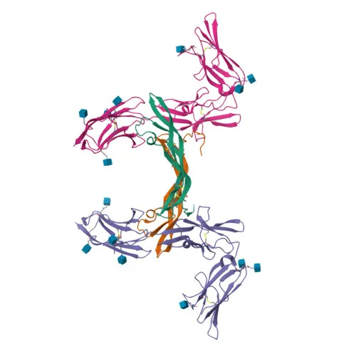
- Platelet-derived growth factor receptor beta (PDGFRB, CD140b) is a cell-surface tyrosine kinase receptor encoded by the PDGFRB gene (Gene ID: 5159). It binds homodimeric PDGFB and PDGFD, as well as PDGFA/PDGFB heterodimers, initiating signaling cascades that regulate embryonic development, cell proliferation, survival, differentiation, chemotaxis, and migration.
- PDGFRB is crucial for vascular development, promoting pericyte and smooth muscle cell recruitment to endothelial cells. It contributes to neointima formation at vascular injury sites and is required for normal cardiovascular system development.
- In the kidney, PDGFRB signaling is essential for mesangial cell recruitment and the formation of a branched capillary network in glomeruli, highlighting its role in organ-specific vascularization.
- Ligand binding triggers autophosphorylation and activation of downstream pathways including PLCG1/PKC, PI3K/AKT1, and RAS/RAF/MAPK (ERK1/2). It also phosphorylates SRC family kinases and adaptor proteins like SHC1, GRB2, CBL, and NCK1, leading to actin cytoskeleton rearrangement and membrane ruffle formation.
- The receptor is localized at the cell membrane, but upon activation, it internalizes into cytoplasmic vesicles and traffics to the lysosome for downregulation. This dynamic subcellular trafficking modulates signal duration and intensity.
- PDGFRB is a member of the type III receptor tyrosine kinase family and shares structural domains including five extracellular immunoglobulin-like domains, a single transmembrane helix, and a split intracellular kinase domain. It can form heterodimers with PDGFRA, which diversifies ligand recognition and signaling output.
- Post-translational modifications include N-linked glycosylation, disulfide bond formation, and ubiquitination. Phosphorylation on multiple tyrosine residues creates docking sites for SH2 domain-containing proteins, a hallmark of signal transduction.
- Clinically, PDGFRB is classified as a proto-oncogene; mutations, overexpression, or chromosomal rearrangements are associated with various malignancies and genetic disorders (e.g., infantile myofibromatosis, Penttinen syndrome). This makes it a valuable biomarker and therapeutic target in oncology.
Experimental Guidance and Technical Tips
- This PE-conjugated rabbit monoclonal antibody is designed for direct flow cytometry analysis of surface CD140b/PDGFRβ on live, unfixed human cells. A matched isotype control is recommended to assess non-specific binding.
- The immunogen spans the extracellular domain (amino acids 33–530), ensuring recognition of the native surface receptor. For optimal staining, titrate the antibody to determine the saturating concentration for your cell type and avoid over-staining.
- If intracellular detection is required, fixation and permeabilization may alter epitope accessibility; validate the staining under appropriate conditions. The 37 kDa recombinant immunogen fragment can serve as a positive control for Western blot or ELISA setups if needed.
- Note that PDGFRB is expressed on various mesenchymal cells, vascular smooth muscle cells, pericytes, and certain tumor cell lines. Include relevant biological controls (e.g., PDGFRB-positive and negative cell lines) in your flow cytometry panel.
CamelBio: Your One-Stop Sourcing Bridge
CamelBio streamlines your IVD development by supplying high-quality raw materials like monoclonal antibodies, validated pairs, and ancillary reagents. As your one-stop sourcing partner, we offer ready-to-use PE-conjugated antibodies targeting PDGFRB to accelerate your flow cytometry-based assays in angiogenesis and cancer research. From bulk antibody orders to rare target sourcing and technical consultation, CamelBio supports your journey from concept to clinic.
Product Datasheet
PE Rabbit anti-Human CD140b/PDGFRβ Monoclonal Antibody for Flow Cytometry - P09619
REQUEST A QUOTE
Our professional team will reply to you within one business day. Please feel free to contact us!