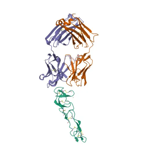
Monoclonal Antibodies
PE Anti-Human CD27 Monoclonal Antibody for Flow Cytometry - P26842
Item Number : CM0006181
Price varies based on specs and customizations
- Application
- FC
- Cross Reactivity
- Human
- Protein Weight
- 29kDa

Shipping:
Contact us to get shipping details Enjoy On-time Dispatch Guarantee.
Why Choose Us
Easy ordering process, quality products, and dedicated support for your business success.
Core Product Specifications and Parameters
| Parameter | Specification |
|---|---|
| Product Name | PE Rabbit anti-Human CD27 mAb |
| Remarks/Alias | T14; S152; Tp55; TNFRSF7; S152. LPFS2 |
| Species | Human |
| Gene ID (Human) | 939 |
| Gene ID | 939 |
| Immunogen | Recombinant protein |
| Source | Rabbit |
| Category | Monoclonal Antibodies |
| Application | FC |
| Cross Reactivity | Human |
| SWISS | P26842 |
| Protein Weight | 29kDa |
| Shipping | Ice bag |
Biological Background: CD27 Function and Localization
- CD27 (also known as TNFRSF7) is a costimulatory immune-checkpoint receptor of the tumor necrosis factor receptor superfamily, expressed on T-cells, NK-cells, and B-cells.
- Binding to its ligand CD70/CD27L triggers CD27-mediated T-cell activation and expansion, which in turn facilitates immune surveillance of B-cells. Related references: PMID:28011863
- Mechanistically, CD70 ligation activates the TRAF2-PTPN6 signaling axis, leading to inhibition of LCK phosphorylation and promoting T-cell memory differentiation. Related references: PMID:38354704
- CD27 also negatively regulates Th2 lymphocyte function in adipose tissue and provides a negative feedback signal to leukocyte differentiation during immune activation.
- CD27 is a single-pass type I membrane protein localized to the cell membrane.
- The protein undergoes post-translational modifications including glycosylation and phosphorylation, and contains structural features such as a signal peptide, transmembrane helix, and disulfide bonds.
- CD27 is predominantly expressed in T-lymphocytes, making it a key marker for T-cell subset analysis.
- The molecular weight of CD27 is approximately 29 kDa (29137 Da according to UniProt).
Experimental Guidance and Technical Tips
- The immunogen corresponds to amino acids 21-192 of human CD27 (NP_001233.2), which spans the extracellular domain, enabling recognition of cell surface CD27.
- For flow cytometry, optimal antibody dilution and staining conditions should be empirically determined using CD27-expressing cells, such as human peripheral blood mononuclear cells (PBMCs) or T-cell lines.
- Consider co-staining with T-cell lineage markers (e.g., CD3, CD4, CD8) to characterize CD27 expression on specific lymphocyte subsets.
- Use appropriate isotype controls and Fc receptor blocking to minimize non-specific binding.
CamelBio: Your One-Stop Sourcing Bridge
CamelBio is your comprehensive IVD raw material sourcing partner, providing validated antibody pairs, optimized monoclonal/polyclonal antibodies, bulk ancillary reagents, and support for rare target development. This PE-conjugated anti-human CD27 antibody supports immunology and immuno-oncology research by enabling detection of the CD27 immune-checkpoint receptor. With an integrated supply chain and technical expertise, CamelBio helps bridge your development needs from concept to clinic.
REQUEST A QUOTE
Our professional team will reply to you within one business day. Please feel free to contact us!