Polyclonal Antibodies
PDHX Rabbit pAb for WB, IP, ELISA - O00330
Item Number : CM0027208
Price varies based on specs and customizations
- Application
- WB, IP, ELISA
- Cross Reactivity
- Human, Mouse, Rat
- Protein Weight
- 54kDa

Shipping:
Contact us to get shipping details Enjoy On-time Dispatch Guarantee.
Why Choose Us
Easy ordering process, quality products, and dedicated support for your business success.
Core Product Specifications and Parameters
| Parameter | Specification |
|---|---|
| Product Name | PDHX Rabbit pAb |
| Remarks/Alias | E3BP; OPDX; PDX1; proX; DLDBP; PDHXD; PDHX |
| Species | Human |
| GeneID (Human) | 8050 |
| GeneID | 8050 |
| Immunogen | Recombinant protein |
| Source | Rabbit |
| Category | Polyclonal Antibodies |
| Application | WB, IP, ELISA |
| Cross Reactivity | Human, Mouse, Rat |
| SWISS | O00330 |
| Protein Weight | 54kDa |
| Shipping | Ice bag |
Biological Background: PDHX Function and Localization
- PDHX, also known as E3-binding protein (E3BP), is a mitochondrial protein that serves as an essential structural component of the pyruvate dehydrogenase complex (PDC).
- Its primary function is to anchor dihydrolipoamide dehydrogenase (E3) to the dihydrolipoamide transacetylase (E2) core, which is critical for the overall enzymatic activity of the PDC.
- The protein contains a lipoyl domain, a characteristic feature of the E2 component of PDC, and is subject to post-translational modifications including acetylation and phosphorylation.
- Alternative splicing of the PDHX gene gives rise to multiple transcript variants, potentially contributing to functional diversity.
- PDHX is localized to the mitochondrial matrix, consistent with its role in oxidative metabolism.
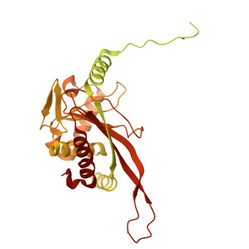
- The protein is part of a macromolecular complex that includes multiple copies of E2 and E3 enzymes, and its structure has been investigated through 3D structural studies.
- Keywords associated with PDHX include "Transit peptide," indicating its mitochondrial targeting, and "Phosphoprotein," highlighting regulatory modification.
- The calculated molecular weight is approximately 54.1 kDa, in agreement with the observed protein weight of 54 kDa.
Experimental Guidance and Technical Tips
- The immunogen corresponds to the N-terminal region (amino acids 1–300) of human PDHX, which may target conserved epitopes in mouse and rat orthologs.
- For Western blot (WB), a band around 54 kDa is expected in mitochondrial lysates from human, mouse, or rat tissues.
- Immunoprecipitation (IP) can be explored for detecting PDHX interactions within the PDC complex.
- ELISA applications may be optimized using recombinant PDHX protein as a standard.
CamelBio: Your One-Stop Sourcing Bridge
At CamelBio, we support diagnostic developers and researchers by offering a wide range of validated antibodies, recombinant proteins, and ancillary reagents. For mitochondrial disease markers like PDHX, we can provide monoclonal alternatives, optimized antigen pairs, and bulk supplies to streamline your assay development. Contact us to source rare targets or discuss your IVD project needs.
REQUEST A QUOTE
Our professional team will reply to you within one business day. Please feel free to contact us!