Polyclonal Antibodies
PDE6G Rabbit pAb for WB, IF/ICC, ELISA - P18545
Item Number : CM0019535
Price varies based on specs and customizations
- Application
- WB, IF/ICC, ELISA
- Cross Reactivity
- Human, Mouse, Rat
- Protein Weight
- 10 kDa

Shipping:
Contact us to get shipping details Enjoy On-time Dispatch Guarantee.
Why Choose Us
Easy ordering process, quality products, and dedicated support for your business success.
Core Product Specifications and Parameters
| Parameter | Value |
|---|---|
| Product Name | PDE6G Rabbit pAb |
| Remarks/Alias | PDEG; RP57; PDE6G |
| Species | Human |
| GeneID (Human) | 5148 |
| GeneID | 5148 |
| Immunogen | Recombinant fusion protein containing a sequence corresponding to amino acids 1-87 of human PDE6G (NP_002593.1). |
| Source | Rabbit |
| Category | Polyclonal Antibodies |
| Application | WB, IF/ICC, ELISA |
| Cross Reactivity | Human, Mouse, Rat |
| SWISS | P18545 |
| Protein Weight | 10 kDa |
| Shipping | Ice bag |
Biological Background: PDE6G Function and Localization
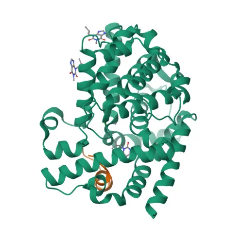
- PDE6G (Phosphodiesterase 6G) is the inhibitory gamma subunit of the rod-specific cGMP phosphodiesterase (PDE6) complex, which plays a central role in the visual phototransduction cascade.
- In the dark, PDE6G binds to the catalytic subunits (PDE6A/PDE6B) to suppress basal cGMP hydrolysis; light-activated transducin (GNAT1) relieves this inhibition, enabling rapid cGMP hydrolysis and signal amplification.
- The PDE6 holoenzyme is a heterotetramer comprising two catalytic and two inhibitory gamma subunits, with PDE6G providing critical regulatory function.
- PDE6G localizes to the photoreceptor outer segment, a modified sensory cilium where phototransduction takes place.
- Post-translational modifications, including acetylation, have been identified in proteomic analyses, suggesting regulatory modulation of PDE6G function.
- Mutations in PDE6G are linked to retinitis pigmentosa, underscoring its essential role in retinal health and vision.
- As a key component of the rod phototransduction pathway, PDE6G is involved in sensory transduction and is categorized under vision-related proteins.
Experimental Guidance and Technical Tips
- Because PDE6G is a low molecular weight protein (~10 kDa), consider using high-percentage SDS-PAGE gels (e.g., 15%) or tricine gels for optimal separation in Western blotting.
- For immunofluorescence/immunocytochemistry (IF/ICC), validate antibody specificity in rod photoreceptor cells or tissues such as retina, where PDE6G is abundantly expressed in outer segments.
- ELISA applications may be suitable for quantifying PDE6G levels in cell or tissue lysates; consider using recombinant protein as a standard for quantitative assays.
- The immunogen consisting of the full-length PDE6G sequence suggests that the antibody may recognize both native and denatured forms, but confirm specificity under the experimental conditions used.
CamelBio: Your One-Stop Sourcing Bridge
CamelBio is a specialized partner for diagnostic manufacturers and research institutes, providing one-stop access to high-quality IVD raw materials and technical services from concept to clinic. This anti-PDE6G antibody supports research in vision, phototransduction, and retinal diseases, contributing to assay development and biomarker studies. With capabilities in supplying validated antibody pairs, optimized recombinant proteins, and ancillary reagents, CamelBio streamlines your sourcing needs for rare targets like PDE6G.
REQUEST A QUOTE
Our professional team will reply to you within one business day. Please feel free to contact us!