Monoclonal Antibodies
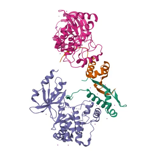
PAK1 Rabbit mAb for WB, IF/ICC, ELISA - Q13153
Item Number : CM0002644
Price varies based on specs and customizations
- Application
- WB, IF/ICC, ELISA
- Cross Reactivity
- Human, Mouse, Rat
- Protein Weight
- 61kDa

Shipping:
Contact us to get shipping details Enjoy On-time Dispatch Guarantee.
Why Choose Us
Easy ordering process, quality products, and dedicated support for your business success.
Core Product Specifications and Parameters
| Parameter | Value |
|---|---|
| Product Name | PAK1 Rabbit mAb |
| Remarks/Alias | IDDMSSD; p65-PAK; PAKalpha; alpha-PAK; PAK1 |
| Species | Human |
| GeneID (human) | 5058 |
| GeneID | 5058 |
| Immunogen | Synthetic Peptide (A synthetic peptide corresponding to a sequence within amino acids 1-100 of human PAK1 (Q13153)) |
| Source | Rabbit |
| Category | Monoclonal Antibodies |
| Application | WB, IF/ICC, ELISA |
| Cross Reactivity | Human, Mouse, Rat |
| SWISS | Q13153 |
| Protein Weight | 61kDa |
| Shipping | Ice bag |
Biological Background: PAK1 Function and Localization
- Encoded by the PAK1 gene, PAK1 (p21-activated kinase 1) is a serine/threonine-protein kinase also known as alpha-PAK, p65-PAK, and PAKalpha. It belongs to the PAK family of kinases that act as effectors for the small GTPases CDC42 and RAC1.
- PAK1 serves as a critical signaling node downstream of integrins and receptor tyrosine kinases, regulating diverse cellular processes including cytoskeleton dynamics, cell adhesion, migration, proliferation, apoptosis, mitosis, and vesicle-mediated transport (PubMed:10551809, PubMed:11896197, PubMed:12876277). Related references: PMID:10551809 PMID:11896197 PMID:12876277
- Upon activation by GTP-bound CDC42 and RAC1, PAK1 links Rho-family GTPases to the JNK MAP kinase pathway and phosphorylates downstream targets such as MAP2K1, RAF1, and LIMK1, thereby influencing gene expression and actin remodeling (PubMed:8805275, PubMed:9528787, PubMed:11733498, PubMed:23633677). Related references: PMID:8805275 PMID:9528787 PMID:11733498 PMID:23633677
- PAK1 localizes to multiple subcellular compartments including the cytoplasm, focal adhesions, lamellipodia, membrane ruffles, invadopodia, nucleoplasm, and centrosomes, consistent with its pleiotropic roles in cell motility, adhesion, and nuclear signaling.
- The kinase plays a role in the reorganization of the actin cytoskeleton and focal adhesion complexes, directly phosphorylating regulators such as myosin light chain and cofilin to modulate actomyosin contractility and cell shape (PubMed:9032240, PubMed:9395435). Related references: PMID:9032240 PMID:9395435
- In neurons, PAK1 is essential for dendritic spine formation, excitatory synapse development, and GABAergic inhibitory transmission, partly through F-actin stabilization and myosin II regulatory light chain phosphorylation.
- PAK1 has been implicated in cancer: it is overexpressed in gastric cancer cells and tissues (PubMed:25766321), promotes cell migration and invasion, and phosphorylates substrates involved in chromatin remodeling (MORC2, PubMed:23260667) and epithelial–mesenchymal transition (SNAI1, PubMed:15833848). Related references: PMID:25766321 PMID:23260667 PMID:15833848
- Additionally, PAK1 participates in insulin secretion (PubMed:22669945), DNA damage response, and stress granule dynamics (PubMed:20417602). Related references: PMID:22669945 PMID:20417602
Experimental Guidance and Technical Tips
- The immunogen is a synthetic peptide spanning amino acids 1-100 of human PAK1, which corresponds to the N-terminal region. This region is likely conserved across human, mouse, and rat, consistent with the reported cross-reactivity.
- For Western blot (WB), the predicted molecular weight of PAK1 is approximately 61 kDa; however, post-translational modifications or splice variants may result in additional bands. Use appropriate reducing conditions and validate with known positive controls (e.g., HeLa or MCF-7 cell lysates).
- In immunofluorescence (IF/ICC), PAK1 exhibits dynamic subcellular distribution, appearing in focal adhesions, membrane ruffles, and the nucleus depending on cell type and stimulation. Fixation with paraformaldehyde and permeabilization with Triton X-100 or methanol may be suitable, but optimization is recommended for your specific cell line and conditions.
- For ELISA, the antibody may be used in direct or indirect formats. Consider using the immunogen peptide as a positive control to establish assay sensitivity and specificity.
- This antibody has not been tested in other applications; if used for IP, ChIP, or functional blocking, thorough validation in the relevant sample system is advised.
CamelBio: Your One-Stop Sourcing Bridge
CamelBio supports diagnostic manufacturers, research laboratories, and IVD developers with comprehensive raw-material solutions—from validated antibody pairs and optimized monoclonal/polyclonal antibodies to bulk ancillary reagents. Whether your work focuses on PAK1 signaling in cancer, neuroscience, or metabolic pathways, our one-stop platform simplifies procurement and accelerates project timelines. For custom antibody development, recombinant protein production, or rare-target sourcing, the CamelBio team is ready to assist with flexible supply options tailored to your clinical or R&D needs.
REQUEST A QUOTE
Our professional team will reply to you within one business day. Please feel free to contact us!