Polyclonal Antibodies
MYOC Rabbit pAb for WB, ELISA - Q99972
Item Number : CM0024287
Price varies based on specs and customizations
- Application
- WB, ELISA
- Cross Reactivity
- Human, Mouse
- Protein Weight
- 57kDa

Shipping:
Contact us to get shipping details Enjoy On-time Dispatch Guarantee.
Why Choose Us
Easy ordering process, quality products, and dedicated support for your business success.
Core Product Specifications and Parameters
| Parameter | Value |
|---|---|
| Product Name | MYOC Rabbit pAb |
| Remarks/Alias | GPOA; JOAG; TIGR; GLC1A; JOAG1; MYOC |
| Species | Human |
| GeneID (Human) | 4653 |
| GeneID | 4653 |
| Immunogen | Recombinant protein |
| Source | Rabbit |
| Category | Polyclonal Antibodies |
| Application | WB, ELISA |
| Cross Reactivity | Human, Mouse |
| SWISS | Q99972 |
| Protein Weight | 57kDa |
| Shipping | Ice bag |
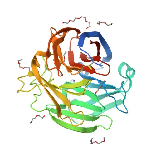
Biological Background: Myocilin Function and Localization
- Myocilin is a secreted glycoprotein also known as the trabecular meshwork-induced glucocorticoid response protein (TIGR) and exists as a 55 kDa subunit, with a full-length molecular weight of approximately 57 kDa.
- It regulates multiple signaling pathways to control cell adhesion, cell-matrix adhesion, cytoskeleton organization, and cell migration, promoting substrate adhesion and focal contact formation while negatively regulating stress fiber assembly via Rho signaling.
- Myocilin modulates the actin cytoskeleton through interactions with Wnt signaling components and promotes cell migration by activating PTK2 and downstream phosphatidylinositol 3-kinase pathways.
- Beyond adhesion, it plays roles in osteoblast differentiation, peripheral nervous system myelination via ERBB2/ERBB3, muscle hypertrophy through the dystrophin-associated protein complex, and positive regulation of mitochondrial depolarization.
- Subcellularly, myocilin is secreted and also localized to the Golgi apparatus, cytoplasmic vesicles, extracellular space and matrix, exosomes, mitochondria (intermembrane space, inner and outer membranes), rough endoplasmic reticulum, cell projections, cilia, and the endoplasmic reticulum.
- It is widely expressed, with high levels in various muscle types, ciliary body, heart, and bone marrow-derived mesenchymal stem cells, and is detected in aqueous humor and the eye, particularly in the trabecular meshwork where increased expression is associated with glaucoma.
- Key post-translational features include glycosylation, palmitoylation, disulfide bonds, and calcium binding, reflecting its association with membranes and secretory pathways.
Experimental Guidance and Technical Tips
- The immunogen corresponds to amino acids 245–504 of human MYOC, covering a region that may detect both full-length and proteolytic fragments; consider this when analyzing bands in WB.
- For WB, optimizing loading amounts and antibody dilution is recommended, as myocilin expression levels can vary significantly between cell types and tissues.
- The antibody is indicated for WB and ELISA; its suitability for other applications (e.g., IHC, IF) has not been assessed and should be validated in the relevant sample system.
- Cross-reactivity is reported for human and mouse; verify reactivity in other species if needed, as the immunogen sequence shows conservation.
CamelBio: Your One-Stop Sourcing Bridge
CamelBio supports IVD manufacturers and researchers with comprehensive access to monoclonal and polyclonal antibodies, validated antibody pairs, and ancillary reagents. For studies on glaucoma, cell adhesion, or myocilin-related signaling, we offer reliable sourcing of rare-target raw materials. Partner with us to streamline your diagnostic development from concept to clinic.
REQUEST A QUOTE
Our professional team will reply to you within one business day. Please feel free to contact us!