Monoclonal Antibodies
Mouse anti-Human CD3 Monoclonal Antibody for Flow Cytometry - P07766
Item Number : CM0010517
Price varies based on specs and customizations
- Application
- FC
- Cross Reactivity
- Human
- Protein Weight
- 23kDa

Shipping:
Contact us to get shipping details Enjoy On-time Dispatch Guarantee.
Why Choose Us
Easy ordering process, quality products, and dedicated support for your business success.
Core Product Specifications and Parameters
| Parameter | Value |
|---|---|
| Product Name | Mouse anti-Human CD3 mAb |
| Remarks/Alias | T3E; TCRE; IMD18; CD3epsilon |
| Species | Human |
| Gene ID (Human) | 916 |
| Immunogen | Recombinant protein of human CD3 |
| Source | Mouse |
| Category | Monoclonal Antibodies |
| Application | FC |
| Cross Reactivity | Human |
| SWISS | P07766 |
| Protein Weight | 23kDa |
| Shipping | Ice bag |
Biological Background: CD3 Epsilon Chain Function and Localization
- CD3E encodes the epsilon chain of the T-cell receptor (TCR)-CD3 complex, a multisubunit receptor essential for T-lymphocyte activation and adaptive immunity. Related references: PMID:15294938 PMID:15546002 PMID:2470098
- Upon TCR engagement, the immunoreceptor tyrosine-based activation motifs (ITAMs) within the cytoplasmic tails of CD3 chains become phosphorylated by Src kinases, enabling downstream signaling. Related references: PMID:2470098 PMID:40592325
- Phosphorylated CD3E ITAMs serve as docking sites for the kinase ZAP70, which is then phosphorylated and converted into an active enzyme, propagating T-cell activation signals.
- CD3E is critical for proper T-cell development, functioning in both pre-TCR and mature TCR complexes during thymocyte selection.
- The cytoplasmic region of CD3E contains endocytosis motifs that mediate internalization and surface down-regulation of TCR-CD3 complexes, modulating T-cell responsiveness. Related references: PMID:10384095 PMID:26507128
- CD3E also functions as a receptor for ITPRIPL1, which inhibits T-cell activation by promoting NCK1 interaction and blocking the ERK-NFkB signaling cascade. Related references: PMID:12110186 PMID:38614099
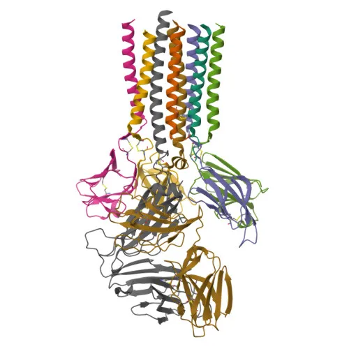
- The protein localizes to the cell membrane as a single-pass type I membrane protein, featuring an extracellular immunoglobulin-like domain and a cytoplasmic ITAM-containing region.
- CD3E undergoes key post-translational modifications, primarily tyrosine phosphorylation, which is central to its signaling function. Related references: PMID:2470098 PMID:40592325
Experimental Guidance and Technical Tips
- The immunogen used is recombinant human CD3 protein; ensure your sample preparation preserves the native CD3 conformation for optimal antibody binding.
- For flow cytometry, perform antibody titration using your specific cell population to determine the optimal staining concentration and minimize background.
- Validate antibody cross-reactivity in your experimental system, as this clone is specific for human CD3 and may not recognize CD3 from other species.
- exhibits excitation at ~633 nm; design your multicolor panel with compatible fluorophores to avoid spectral overlap.
- CD3 is expressed on mature T lymphocytes; use appropriate gating strategies and viability dyes to identify live T-cell subsets accurately.
CamelBio: Your One-Stop Sourcing Bridge
CamelBio connects diagnostic manufacturers and research laboratories with a comprehensive portfolio of IVD raw materials, including optimized monoclonal antibodies like this anti-CD3 clone. Whether you require validated antibody pairs, bulk ancillary reagents, or rare-target sourcing for T-cell immunodetection, we provide end-to-end support from concept to clinical application. Contact our team to streamline your procurement and accelerate assay development.
REQUEST A QUOTE
Our professional team will reply to you within one business day. Please feel free to contact us!