Monoclonal Antibodies
MICA Rabbit mAb for WB, ELISA - Q29983
Item Number : CM0003447
Price varies based on specs and customizations
- Application
- WB, ELISA
- Cross Reactivity
- Human, Mouse
- Protein Weight
- 43kDa

Shipping:
Contact us to get shipping details Enjoy On-time Dispatch Guarantee.
Why Choose Us
Easy ordering process, quality products, and dedicated support for your business success.
Core Product Specifications and Parameters
| Parameter | Value |
|---|---|
| Product Name | MICA Rabbit mAb |
| Remarks/Alias | MIC-A; PERB11.1; MICA |
| Species | Human |
| GeneID (Human) | 100507436 |
| GeneID | 100507436 |
| Immunogen | Synthetic peptide |
| Source | Rabbit |
| Category | Monoclonal Antibodies |
| Application | WB, ELISA |
| Cross Reactivity | Human, Mouse |
| SWISS | Q29983 |
| Protein Weight | 43kDa |
| Shipping | Ice bag |
Biological Background: MICA Function and Localization
- MICA (MHC class I polypeptide-related sequence A) is a non-classical MHC class I molecule, also designated MIC-A or PERB11.1.
- It acts as a ligand for the NKG2D receptor, which is present on NK cells, γδ T cells, and CD8+ αβ T cells. Related references: PMID:11491531, PMID:11777960
- Under cellular stress (e.g., infection, DNA damage), MICA is upregulated, and NKG2D engagement triggers cytotoxicity and cytokine production. Related references: PMID:10426993
- MICA is broadly expressed in tissues such as gastric epithelium, monocytes, keratinocytes, endothelial cells, and fibroblasts, but absent from the central nervous system. It is frequently overexpressed in epithelial tumors (lung, breast, kidney, ovary, prostate, colon), where it associates with increased γδ T-cell infiltration.
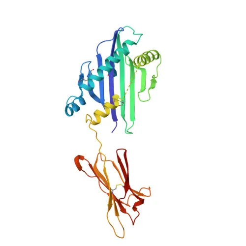
- Subcellularly, MICA localizes to the cell membrane and cytoplasm; it is a transmembrane glycoprotein with a signal peptide, extracellular Ig-like domains, a transmembrane helix, and a cytoplasmic tail.
- Post-translational modifications include N-linked glycosylation and S-palmitoylation; the protein also contains a conserved disulfide bond.
- Alternative splicing generates multiple isoforms, and MICA is implicated in host-virus interactions and ubiquitination pathways.
Experimental Guidance and Technical Tips
- The immunogen (aa 50-150) targets the extracellular domain, which may favor detection of both surface and shed soluble forms of MICA.
- For western blot, a band around 43 kDa is expected; consider using reducing conditions and validating detection in MICA-expressing tumor cell lines (e.g., HCT-116, HeLa).
- In ELISA applications, this monoclonal antibody could be used as a capture or detection reagent; pairing with a different clone may enable sandwich ELISA development. Validate specificity with recombinant MICA standard.
CamelBio: Your One-Stop Sourcing Bridge
CamelBio provides diagnostic manufacturers, research institutes, and laboratories with one-stop access to validated IVD raw materials, technical services, and supply chain consulting. For MICA-related immuno-oncology and cellular stress research, we offer optimized monoclonal and polyclonal antibodies, bulk ancillary reagents, and support for custom raw-material procurement. From concept to clinic, CamelBio streamlines your development pipeline with integrated sourcing and expert guidance.
REQUEST A QUOTE
Our professional team will reply to you within one business day. Please feel free to contact us!