Polyclonal Antibodies
MADCAM1 Rabbit pAb for WB, IHC-P, ELISA - Q13477
Item Number : CM0027991
Price varies based on specs and customizations
- Application
- WB, IHC-P, ELISA
- Cross Reactivity
- Human, Mouse, Rat
- Protein Weight
- 40kDa

Shipping:
Contact us to get shipping details Enjoy On-time Dispatch Guarantee.
Why Choose Us
Easy ordering process, quality products, and dedicated support for your business success.
Core Product Specifications and Parameters
| Parameter | Value |
|---|---|
| Product Name | MADCAM1 Rabbit pAb |
| Remarks/Alias | MACAM1; MADCAM1 |
| Species | Human |
| Gene ID (Human) | 8174 |
| Gene ID | 8174 |
| Immunogen | A synthetic peptide corresponding to a sequence within amino acids 100-200 of human MADCAM1 (NP_570116.2). |
| Source | Rabbit |
| Category | Polyclonal Antibodies |
| Application | WB, IHC-P, ELISA |
| Cross Reactivity | Human, Mouse, Rat |
| SWISS | Q13477 |
| Protein Weight | 40kDa |
| Shipping | Ice bag |
Biological Background: MADCAM1 Function and Localization
- MADCAM1 (mucosal addressin cell adhesion molecule 1) is a cell adhesion receptor expressed on mucosal venules, directing lymphocyte trafficking into mucosal tissues such as Peyer’s patches and intestinal lamina propria.
- It binds both integrin α4β7 and L-selectin, thereby regulating leukocyte passage and retention. Isoform 2, which lacks the mucin-like domain, may specialize in integrin α4β7‑dependent adhesion strengthening independent of L-selectin.
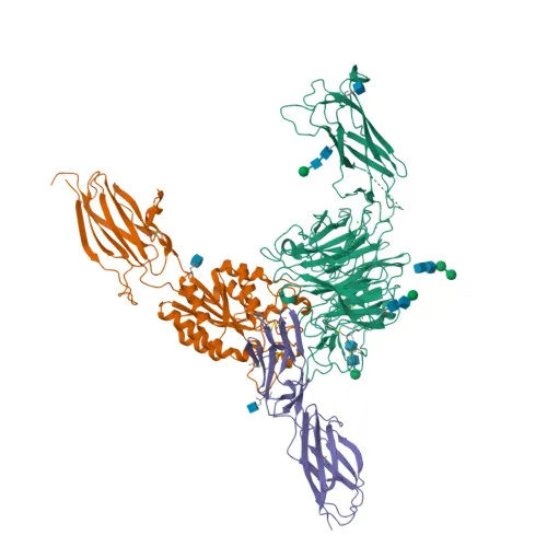
- The protein is a type I transmembrane glycoprotein belonging to the immunoglobulin superfamily, containing immunoglobulin-like domains and a mucin-rich region (in the canonical isoform).
- Subcellular localization is predominantly at the plasma membrane, consistent with its role in cell–cell interactions.
- Tissue specificity: Highly expressed on high endothelial venules (HEV) and lamina propria venules in the small intestine; lower expression in colon and spleen. Very low in pancreas and brain; absent in thymus, prostate, ovaries, testis, heart, placenta, lung, liver, skeletal muscle, kidney, and peripheral blood leukocytes.
- Post‑translational modifications include N‑linked glycosylation and disulfide bonds, essential for structural integrity and ligand binding. Keywords also highlight alternative splicing and 3D‑structure availability.
Experimental Guidance and Technical Tips
- The immunogen spans amino acids 100‑200, an extracellular region; consider that the antibody may recognize both native and denatured MADCAM1, but epitope accessibility in fixed tissues should be validated.
- For Western blot, a band around 40 kDa is expected under reducing conditions; use appropriate molecular weight markers and verify with a positive control (e.g., human intestinal tissue lysate).
- In IHC‑P, antigen retrieval (heat‑ or enzyme‑based) may be necessary to expose the epitope; test multiple retrieval methods and antibody dilutions on known positive and negative tissues.
- ELISA conditions such as coating buffer and detection system should be optimized individually; a peptide competition assay can help confirm specificity.
- Cross‑reactivity is reported for human, mouse, and rat; for other species, perform sequence alignment and preliminary testing before application.
CamelBio: Your One-Stop Sourcing Bridge
CamelBio is your dedicated partner for IVD raw materials, providing one‑stop access to validated antibodies like MADCAM1 for diagnostic and research applications. We support your mucosal immunology projects with ready‑to‑use polyclonal antibodies, custom antibody development, and bulk ordering options. Whether you need optimized antibody pairs, ancillary reagents, or rare‑target sourcing, CamelBio streamlines your procurement from concept to clinic.
REQUEST A QUOTE
Our professional team will reply to you within one business day. Please feel free to contact us!