Polyclonal Antibodies
MAD1/MAD1L1 Rabbit Polyclonal Antibody for WB, IF/ICC, ELISA - Q9Y6D9
Item Number : CM0014408
Price varies based on specs and customizations
- Application
- WB, IF/ICC, ELISA
- Cross Reactivity
- Human
- Protein Weight
- 83kDa

Shipping:
Contact us to get shipping details Enjoy On-time Dispatch Guarantee.
Why Choose Us
Easy ordering process, quality products, and dedicated support for your business success.
Core Product Specifications and Parameters
| Parameter | Value |
|---|---|
| Product Name | MAD1/MAD1L1 Rabbit pAb |
| Remarks/Alias | MAD1; MVA7; PIG9; TP53I9; TXBP181; MAD1/MAD1L1 |
| Species | Human |
| GeneID (human) | 8379 |
| GeneID | 8379 |
| Immunogen | Recombinant fusion protein containing a sequence corresponding to amino acids 360-700 of human MAD1L1 (NP_001013859.1) |
| Source | Rabbit |
| Category | Polyclonal Antibodies |
| Application | WB, IF/ICC, ELISA |
| Cross Reactivity | Human |
| SWISS | Q9Y6D9 |
| Protein Weight | 83kDa |
| Shipping | Ice bag |
Biological Background: MAD1 Function and Localization
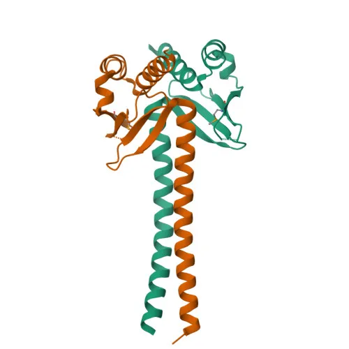
- MAD1L1 encodes the mitotic spindle assembly checkpoint protein MAD1, also known as mitotic arrest deficient 1-like protein 1, HsMAD1, hMAD1, and Tax-binding protein 181.
- MAD1 is a core component of the spindle-assembly checkpoint (SAC), which prevents anaphase onset until all chromosomes are correctly attached to spindle microtubules at the metaphase plate. Related references: PMID:10049595, PMID:20133940, PMID:29162720
- At unattached kinetochores, MAD1 forms a heterotetrameric complex with closed-conformation MAD2L1 (C-MAD2) and catalyzes the conformational conversion of open MAD2L1 (O-MAD2) to C-MAD2, a key step in generating the diffusible "wait-anaphase" signal. Related references: PMID:10049595, PMID:20133940, PMID:29162720
- In the cytoplasm, MAD1 can sequester MAD2L1, impairing SAC function and contributing to chromosomal instability in hepatocellular carcinomas.
- Subcellular localization includes the nucleus, kinetochores, centrosomes, spindle poles, and cytoplasm, reflecting its dynamic role during mitosis.
- Tissue specificity: MAD1 is expressed in hepatocellular carcinomas and hepatoma cell lines at the protein level.
- Post-translational modifications: MAD1 undergoes phosphorylation, acetylation, and ubiquitin-like conjugation, which may regulate its stability and activity.
Experimental Guidance and Technical Tips
- The immunogen corresponds to the central region (aa 360–700) of human MAD1L1, which may allow recognition of both full-length and possible cleavage products; consider this when analyzing band patterns in WB.
- For WB, the predicted molecular weight is ~83 kDa; use appropriate positive controls such as HeLa cell lysates or other actively dividing cell lines to confirm specificity.
- For IF/ICC, permeabilization is required to access nuclear and kinetochore-localized MAD1; validate staining in the relevant sample system under mitotic arrest conditions (e.g., nocodazole treatment).
- ELISA performance may depend on coating buffer and antigen presentation; consider standard indirect ELISA protocols with recombinant MAD1 protein as a reference.
CamelBio: Your One-Stop Sourcing Bridge
CamelBio provides diagnostic manufacturers and researchers with reliable access to critical IVD raw materials. For cell cycle and oncology research, we offer validated antibodies against checkpoint proteins like MAD1, alongside matching antigen standards and ancillary reagents to accelerate assay development. Our sourcing network ensures consistent supply of high-quality polyclonal antibodies and supports from concept to clinical application.
Product Datasheet
MAD1/MAD1L1 Rabbit Polyclonal Antibody for WB, IF/ICC, ELISA - Q9Y6D9
REQUEST A QUOTE
Our professional team will reply to you within one business day. Please feel free to contact us!