Polyclonal Antibodies
LC3B Rabbit Polyclonal Antibody for WB, IHC-P, IF/ICC, ELISA - Q9GZQ8
Item Number : CM0017964
Price varies based on specs and customizations
- Application
- WB, IHC-P, IF/ICC, ELISA
- Cross Reactivity
- Human, Mouse, Rat
- Protein Weight
- 15kDa

Shipping:
Contact us to get shipping details Enjoy On-time Dispatch Guarantee.
Why Choose Us
Easy ordering process, quality products, and dedicated support for your business success.
Core Product Specifications and Parameters
| Parameter | Description |
|---|---|
| Product Name | LC3B Rabbit pAb |
| Remarks/Alias | LC3B; ATG8F; MAP1LC3B-a; MAP1A/1BLC3 |
| Species | Human |
| Gene ID | 81631 |
| Immunogen | Recombinant fusion protein containing a sequence corresponding to amino acids 1-100 of human LC3B (NP_073729.1) |
| Source | Rabbit |
| Category | Polyclonal Antibodies |
| Application | WB, IHC-P, IF/ICC, ELISA |
| Cross Reactivity | Human, Mouse, Rat |
| SWISS | Q9GZQ8 |
| Protein Weight | 15kDa |
| Shipping | Ice bag |
Biological Background: LC3B Function and Localization
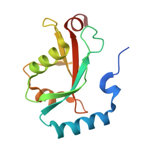
- LC3B (Microtubule-associated protein 1 light chain 3 beta), also known as ATG8F or MAP1LC3B, is a core component of the autophagy pathway and belongs to the ATG8 ubiquitin-like protein family.
- It functions as a ubiquitin-like modifier crucial for the elongation of phagophore membranes during autophagosome formation. Related references: PMID:20418806, PMID:23209295, PMID:28017329
- LC3B plays a pivotal role in selective autophagy subtypes such as mitophagy, where it anchors autophagolysosomes to outer mitochondrial membranes via C-18 ceramide binding after mitochondrial fission. Related references: PMID:22922758
- It promotes primary ciliogenesis by mediating the autophagic removal of OFD1 from centriolar satellites. Related references: PMID:24089205
- Upon nutrient stress, LC3B interacts with reticulophagy receptor TEX264 to remodel ER subdomains into autophagosomes, facilitating ER turnover. Related references: PMID:31006537, PMID:31006538
- LC3B directly recruits the actin nucleation factor JMY to phagophore membranes, stimulating actin polymerization and autophagosome biogenesis. Related references: PMID:30420355
- Subcellularly, LC3B is found on autophagosome membranes, cytoplasmic vesicles, mitochondrial membranes, and the cytoskeleton; it undergoes a characteristic lipidation shift from cytosolic LC3B-I to membrane-bound LC3B-II.
- Tissue expression is highest in heart, brain, skeletal muscle, and testis, with lower levels in liver.
Experimental Guidance and Technical Tips
- The immunogen (amino acids 1-100 of human LC3B) lies within the N-terminal region; consider confirming antibody specificity using LC3B knockout or knockdown models.
- For Western blotting, LC3B typically appears as two bands: a cytosolic ~15 kDa LC3B-I and a faster-migrating ~14 kDa LC3B-II (lipidated form) under reducing conditions; both forms should be considered when analyzing autophagy flux.
- In immunofluorescence applications, fixation and permeabilization protocols may need optimization to reveal punctate autophagosome staining under autophagy-inducing conditions (e.g., starvation or pharmacological treatment).
- For ELISA, ensure proper coating buffer and antibody dilution; validate against known autophagy markers in the relevant sample system.
CamelBio: Your One-Stop Sourcing Bridge
As a dedicated IVD raw‑material partner, CamelBio connects diagnostic manufacturers and research labs with rigorously developed antibodies like this LC3B polyclonal antibody. We support your autophagy‑related assay development with validated antibody pairs, optimized monoclonal and polyclonal antibodies, bulk ancillary reagents, and expert sourcing for rare‑target raw materials—helping streamline your path from concept to clinic.
Product Datasheet
LC3B Rabbit Polyclonal Antibody for WB, IHC-P, IF/ICC, ELISA - Q9GZQ8
REQUEST A QUOTE
Our professional team will reply to you within one business day. Please feel free to contact us!