Polyclonal Antibodies
KO Validated CDK7 Rabbit Polyclonal Antibody for WB, IF/ICC, ELISA - P50613
Item Number : CM0017844
Price varies based on specs and customizations
- Application
- WB, IF/ICC, ELISA
- Cross Reactivity
- Human, Mouse, Rat
- Protein Weight
- 39kDa

Shipping:
Contact us to get shipping details Enjoy On-time Dispatch Guarantee.
Why Choose Us
Easy ordering process, quality products, and dedicated support for your business success.
Core Product Specifications and Parameters
| Parameter | Value |
|---|---|
| Product Name | [KO Validated] CDK7 Rabbit pAb |
| Remarks/Alias | CAK; CAK1; HCAK; MO15; STK1; CDKN7; p39MO15; K7 |
| Species | Human |
| Gene ID (Human) | 1022 |
| Gene ID | 1022 |
| Immunogen | Recombinant protein|Recombinant fusion protein containing a sequence corresponding to amino acids 211-346 of human CDK7 (NP_001790.1). |
| Source | Rabbit |
| Category | Polyclonal Antibodies |
| Application | WB, IF/ICC, ELISA |
| Cross Reactivity | Human, Mouse, Rat |
| SWISS | P50613 |
| Protein Weight | 39kDa |
| Shipping | Ice bag |
Biological Background: CDK7 Function and Localization
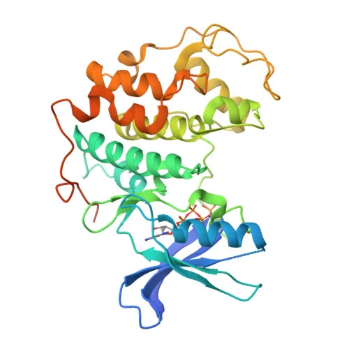
- Cyclin-dependent kinase 7 (CDK7) is a serine/threonine kinase that serves as the catalytic subunit of the CDK-activating kinase (CAK) complex, playing a pivotal role in both cell cycle regulation and RNA polymerase II–mediated transcription. Related references: PMID:9852112
- CDK7 phosphorylates the C-terminal domain (CTD) of RNA polymerase II (POLR2A) at Ser5, a critical step for transcription initiation and promoter escape. Related references: PMID:19136461, PMID:26257281, PMID:28768201
- As part of the CAK complex, CDK7 activates major cyclin-dependent kinases (CDK1, CDK2, CDK4, CDK6) by phosphorylating their T-loop threonines, thereby driving cell cycle progression. Related references: PMID:41100585
- CDK7 is localized in both the nucleus and the cytoplasm, with enrichment in the perinuclear region.
- It is ubiquitously expressed across tissues, consistent with its fundamental roles in cell division and gene expression.
- In response to DNA damage, CDK7 phosphorylates and activates p53, which in turn inhibits CDK7, forming a feedback loop that can lead to cell cycle arrest or apoptosis. Related references: PMID:9372954, PMID:9840937
- CDK7 also phosphorylates other substrates including SPT5, SF1, and CDK11B, linking it to additional regulatory networks. Related references: PMID:9372954
- Key post-translational modifications of CDK7 include acetylation and phosphorylation, which modulate its activity and interactions.
Experimental Guidance and Technical Tips
- The immunogen spans amino acids 211–346 within the kinase domain, which may help detect full-length CDK7 (~39 kDa) and potential isoforms; consider using the recombinant protein as a positive control.
- For Western blotting, validate the antibody in cell lines such as HeLa or HEK293 and use appropriate lysis buffers to preserve phosphorylation states.
- In immunofluorescence/ICC, expect nuclear and perinuclear staining patterns; include isotype controls and confirm specificity in CDK7‑depleted cells where possible.
- Cross-reactivity with mouse and rat CDK7 is reported; validate in relevant tissue lysates if these species are part of your experimental system.
- For ELISA applications, optimize coating conditions and consider using the immunogen or purified CDK7 protein as a standard.
CamelBio: Your One-Stop Sourcing Bridge
CamelBio empowers diagnostic manufacturers and research laboratories with efficient access to high-quality IVD raw materials. This CDK7 polyclonal antibody highlights our focus on key targets in cell cycle and transcriptional regulation. Our integrated supply network offers validated antibody pairs, optimized polyclonal and monoclonal antibodies, bulk ancillary reagents, and custom sourcing for rare targets to accelerate your assay development and production.
Product Datasheet
KO Validated CDK7 Rabbit Polyclonal Antibody for WB, IF/ICC, ELISA - P50613
REQUEST A QUOTE
Our professional team will reply to you within one business day. Please feel free to contact us!