Polyclonal Antibodies
KO Validated Anti-PDCD6 Polyclonal Antibody (ALG2) for WB, IP, ELISA - O75340
Item Number : CM0025571
Price varies based on specs and customizations
- Application
- WB, IP, ELISA
- Cross Reactivity
- Human, Mouse, Rat
- Protein Weight
- 22kDa

Shipping:
Contact us to get shipping details Enjoy On-time Dispatch Guarantee.
Why Choose Us
Easy ordering process, quality products, and dedicated support for your business success.
Core Product Specifications and Parameters
| Parameter | Value |
|---|---|
| Product Name | [KO Validated] PDCD6 Rabbit pAb |
| Remarks/Alias | ALG2; ALG-2; PEF1B; D6 |
| Species | Human |
| Gene ID (Human) | 10016 |
| Immunogen | Recombinant fusion protein containing a sequence corresponding to amino acids 1-191 of human PDCD6 (NP_037364.1). |
| Source | Rabbit |
| Category | Polyclonal Antibodies |
| Application | WB, IP, ELISA |
| Cross Reactivity | Human, Mouse, Rat |
| SWISS | O75340 |
| Protein Weight | 22kDa |
| Shipping | Ice bag |
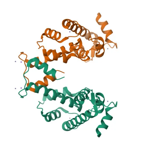
Biological Background: PDCD6 Function and Localization
- PDCD6 (programmed cell death protein 6, alias ALG-2) is a calcium-binding protein of the penta-EF-hand family that functions as a calcium sensor in ER-Golgi vesicular transport, endosomal biogenesis, and membrane repair by bridging unrelated proteins in response to calcium, triggering membrane translocation. Related references: PMID:20691033, PMID:25667979
- It facilitates ER-Golgi transport by promoting the association between PDCD6IP and TSG101, thereby bridging the ESCRT-III and ESCRT-I complexes. Related references: PMID:19520058
- Together with PEF1, PDCD6 acts as a calcium-dependent adapter for the BCR(KLHL12) complex, regulating COPII coat size and collagen export, a process essential for neural crest specification. Related references: PMID:27716508
- In the endosomal pathway, PDCD6 regulates the distribution and function of MCOLN1. Related references: PMID:19864416
- It promotes localization and polymerization of TFG at endoplasmic reticulum exit sites. Related references: PMID:27813252
- PDCD6 is required for T-cell receptor-, Fas-, and glucocorticoid-induced apoptosis; interaction with DAPK1 accelerates apoptotic cell death by increasing caspase-3 activity. Related references: PMID:16132846
- Subcellular localization includes endoplasmic reticulum membrane, COPII-coated vesicle membrane, cytoplasm, nucleus, and endosomes.
- Keywords from UniProt: 3D-structure, acetylation (post-translational modification), alternative splicing, angiogenesis, apoptosis, calcium, magnesium, metal-binding, proteomics identification, reference proteome, repeat.
Experimental Guidance and Technical Tips
- This polyclonal antibody, raised against full-length human PDCD6, targets multiple epitopes and is suitable for detecting endogenous levels by Western blot at approximately 22 kDa; validate in the relevant sample system.
- KO validation indicates specificity; consider using appropriate knockout or knockdown controls to confirm target identity.
- Cross-reactivity with mouse and rat has been demonstrated; optimize dilution and conditions when applying to these species.
- For immunoprecipitation and ELISA, perform pre-adsorption and titration experiments to ensure signal specificity, as recommended for polyclonal antibodies.
CamelBio: Your One-Stop Sourcing Bridge
CamelBio provides diagnostic manufacturers, labs, and research institutes with one-stop access to IVD raw materials, including validated antibody pairs, optimized monoclonal and polyclonal antibodies, and bulk ancillary reagents. For your apoptosis and calcium signaling research, we supply high-quality PDCD6 antibodies and can assist with rare target raw-material sourcing, covering every stage from concept to clinic. Partner with us for comprehensive procurement solutions tailored to your development needs.
Product Datasheet
KO Validated Anti-PDCD6 Polyclonal Antibody (ALG2) for WB, IP, ELISA - O75340
REQUEST A QUOTE
Our professional team will reply to you within one business day. Please feel free to contact us!