Monoclonal Antibodies
KD Validated Anti-RHOT1/MIRO1 Rabbit Monoclonal Antibody for WB and ELISA - Q8IXI2
Item Number : CM0007289
Price varies based on specs and customizations
- Application
- WB, ELISA
- Cross Reactivity
- Human, Mouse, Rat
- Protein Weight
- 71kDa

Shipping:
Contact us to get shipping details Enjoy On-time Dispatch Guarantee.
Why Choose Us
Easy ordering process, quality products, and dedicated support for your business success.
Core Product Specifications and Parameters
| Parameter | Value |
|---|---|
| Product Name | [KD Validated] RHOT1/MIRO1 Rabbit mAb |
| Remarks/Alias | ARHT1; MIRO1; MIRO-1; [KO/KD Validated] RHOT1/MIRO1 |
| Species | Human |
| GeneID (human) | 55288 |
| GeneID | 55288 |
| Immunogen | Recombinant fusion protein containing a sequence corresponding to amino acids 310-590 of human RHOT1/MIRO1 (NP_060777.3) |
| Source | Rabbit |
| Category | Monoclonal Antibodies |
| Application | WB, ELISA |
| Cross Reactivity | Human, Mouse, Rat |
| SWISS | Q8IXI2 |
| Protein Weight | 71kDa |
| Shipping | Ice bag |
Biological Background: RHOT1/MIRO1 Function and Localization
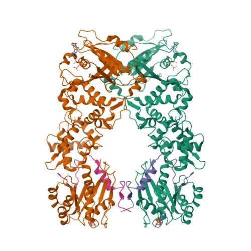
- RHOT1/MIRO1, also designated ARHT1, is a member of the Ras superfamily of small GTPases, specifically an atypical mitochondrial Rho GTPase 1.
- It functions as an NTPase capable of hydrolyzing GTP, ATP, and UTP, a property essential for its role in mitochondrial dynamics. Related references: PMID:30513825
- RHOT1/MIRO1 is a key regulator of mitochondrial trafficking, mediating anterograde transport and controlling the subcellular distribution of mitochondria. Related references: PMID:12482879, PMID:16630562, PMID:22396657
- Under high calcium conditions, RHOT1/MIRO1 promotes mitochondrial fission, linking calcium signaling to mitochondrial morphology. Related references: PMID:27716788
- The protein is anchored to the mitochondrial outer membrane via a single C-terminal transmembrane domain.
- It is subject to post-translational modifications including acetylation and ubiquitination, as indicated by proteomics studies.
- RHOT1/MIRO1 is ubiquitously expressed, with highest levels observed in heart and skeletal muscle.
- Alternative splicing of the RHOT1 gene generates multiple isoforms, which may have tissue-specific or functional differences.
Experimental Guidance and Technical Tips
- The immunogen corresponds to the C-terminal region (aa 310–590) of human RHOT1, covering part of the GTPase domain and the tail; it is expected to recognize the full-length protein.
- For western blot, a predominant band at ~71 kDa is observed in human, mouse, and rat lysates; always validate expression in your experimental system.
- In ELISA applications, the recombinant immunogen fragment can be used as a positive control to assess antibody binding.
- As a rabbit monoclonal antibody, it offers batch-to-batch consistency; optimize working dilutions for your specific assay conditions.
CamelBio: Your One-Stop Sourcing Bridge
CamelBio provides diagnostic manufacturers and research institutions with comprehensive access to IVD raw materials, technical services, and consulting—from concept to clinic. This anti-RHOT1/MIRO1 monoclonal antibody supports research into mitochondrial trafficking and dynamics. Our sourcing capabilities include validated antibody pairs, optimized polyclonal and monoclonal antibodies, bulk ancillary reagents, and customized procurement of rare-target raw materials to accelerate your development pipeline.
REQUEST A QUOTE
Our professional team will reply to you within one business day. Please feel free to contact us!