Polyclonal Antibodies
IRAK3 Rabbit Polyclonal Antibody for WB and ELISA - Q9Y616
Item Number : CM0013864
Price varies based on specs and customizations
- Application
- WB, ELISA
- Cross Reactivity
- Human, Mouse, Rat
- Protein Weight
- 68kDa

Shipping:
Contact us to get shipping details Enjoy On-time Dispatch Guarantee.
Why Choose Us
Easy ordering process, quality products, and dedicated support for your business success.
Core Product Specifications and Parameters
| Parameter | Value |
|---|---|
| Product Name | IRAK3 Rabbit pAb |
| Remarks/Alias | ASRT5; IRAKM; IRAK3 |
| Species | Human |
| GeneID (Human) | 11213 |
| GeneID | 11213 |
| Immunogen | Recombinant fusion protein containing a sequence corresponding to amino acids 367-596 of human IRAK3 (NP_009130.2). |
| Source | Rabbit |
| Category | Polyclonal Antibodies |
| Application | WB, ELISA |
| Cross Reactivity | Human, Mouse, Rat |
| SWISS | Q9Y616 |
| Protein Weight | 68kDa |
| Shipping | Ice bag |
Biological Background: IRAK3 Function and Localization
- IRAK3 (Interleukin-1 receptor-associated kinase 3), also known as IRAK-M, is a member of the IRAK family and exists in multiple isoforms due to alternative splicing.
- It functions as a negative regulator of IL-1 and Toll-like receptor (TLR) signaling pathways (PubMed:10383454, PubMed:29686383). Related references: PMID:10383454 PMID:29686383
- In the TLR signaling complex, IRAK3 inhibits the phosphorylation and dissociation of IRAK1 and IRAK4, thereby stabilizing the receptor complex and dampening downstream signaling (By similarity).
- Upon IL33-induced lung inflammation, IRAK3 positively regulates the expression of cytokines IL6, CSF3, CXCL2, and CCL5 in dendritic cells (PubMed:29686383). Related references: PMID:29686383
- IRAK3 localizes to both the cytoplasm and the nucleus.
- It is predominantly expressed in peripheral blood lymphocytes, and its protein is detected in eosinophils, dendritic cells, and monocytes (PubMed:10383454). Related references: PMID:10383454
- Post-translational modifications include phosphorylation, and the protein contains a disulfide bond and an ATP-binding domain, although its kinase activity is predicted to be inactive.
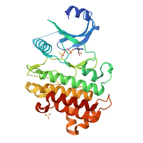
- Structural studies have characterized its inactive kinase domain, providing insights into its regulatory role.
Experimental Guidance and Technical Tips
- The immunogen covers amino acids 367–596 of human IRAK3, corresponding to the C-terminal region containing part of the kinase domain. This region is highly conserved across human, mouse, and rat, supporting the observed cross-reactivity.
- For Western blotting, a band of approximately 68 kDa is expected. Consider using whole cell lysates from human peripheral blood mononuclear cells (PBMCs) or mouse/rat macrophage cell lines (e.g., RAW 264.7) to detect endogenous IRAK3.
- For ELISA applications, this polyclonal antibody may be used as a capture or detection reagent; optimization of coating concentrations and blocking conditions is recommended.
- Cross-reactivity with human, mouse, and rat has been verified; validation in other species or specific tissues should be performed independently.
CamelBio: Your One-Stop Sourcing Bridge
CamelBio provides comprehensive IVD raw material supply solutions, from validated antibody pairs and optimized polyclonal/monoclonal antibodies to bulk ancillary reagents and rare target sourcing. This IRAK3 antibody supports research into innate immunity and inflammatory signaling pathways, and our team can assist in securing additional complementary reagents for your diagnostic development needs. Contact us to streamline your procurement from concept to clinic.
REQUEST A QUOTE
Our professional team will reply to you within one business day. Please feel free to contact us!