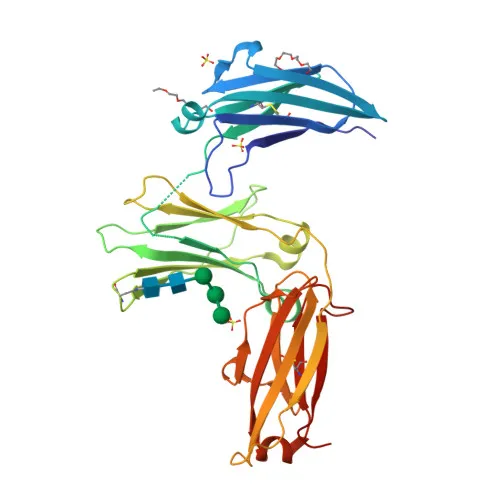
Monoclonal Antibodies
Human IgE Rabbit mAb for WB, FC, ELISA - P01854
Item Number : CM0006982
Price varies based on specs and customizations
- Application
- WB, FC, ELISA
- Cross Reactivity
- Human
- Protein Weight
- 47kDa

Shipping:
Contact us to get shipping details Enjoy On-time Dispatch Guarantee.
Why Choose Us
Easy ordering process, quality products, and dedicated support for your business success.
Core Product Specifications and Parameters
| Parameter | Description |
|---|---|
| Product Name | Human IgE Rabbit mAb |
| Remarks/Alias | Igh2; IGHE; IGHEP1; Human IgE |
| Species | Human |
| Gene ID | 3497 (Human) |
| Immunogen | Recombinant fusion protein containing a sequence corresponding to amino acids 104-428 of human IgE (P01854). |
| Source | Rabbit |
| Category | Monoclonal Antibodies |
| Application | WB, FC, ELISA |
| Cross Reactivity | Human |
| SWISS | P01854 |
| Protein Weight | 47kDa |
| Shipping | Ice bag |
Biological Background: IgE Function and Localization
- IgE is an immunoglobulin heavy chain constant epsilon region that defines the IgE isotype. It is a glycoprotein with intra- and interchain disulfide bonds.
- The antigen-binding specificity of IgE is determined by the variable domains generated through V(D)J recombination and somatic hypermutation, enabling high-affinity recognition. Related references: PMID:17576170, PMID:20176268
- Secreted IgE mediates allergic and anti-parasitic responses through binding to the high-affinity receptor FCER1A on mast cells, basophils, and eosinophils, triggering degranulation and release of histamine, leukotrienes, and cytokines. Related references: PMID:25629393, PMID:33840121, PMID:8114916, PMID:8551243
- IgE also interacts with the low-affinity receptor FCER2 on macrophages to stimulate parasite killing and on B cells to enhance antigen presentation. Related references: PMID:7544003, PMID:2167225
- Membrane-bound IgE exists as long and short isoforms, forming the B cell receptor (BCR) complex. Antigen cross-linking of the BCR controls IgE+ B cell survival and differentiation.
- IgE expression is predominantly found in B lymphocytes upon stimulation with IL-4 and CD40, linking it to Th2-type immune responses.
- Alternative splicing of the IGHE gene generates secreted and two membrane-bound isoforms, each with distinct signaling properties.
- Dysregulated IgE responses contribute to allergic and anaphylactic conditions. Related references: PMID:25629393, PMID:33840121, PMID:7544003
Experimental Guidance and Technical Tips
- The immunogen corresponds to amino acids 104-428 of the constant region of human IgE, covering the CH2-CH4 domains. This design ensures recognition of the Fc region shared by secreted and membrane-bound isoforms.
- For Western blot (WB), consider using reducing conditions; the observed molecular weight may be ~47 kDa as indicated, but glycosylation can shift the band or produce a smear.
- In flow cytometry (FC), this antibody is suitable for detecting membrane-bound IgE on B cells or IgE captured by Fc receptors on basophils/mast cells; proper isotype controls and Fc-blocking steps are recommended.
- For ELISA applications, this monoclonal antibody can serve as a capture or detection reagent for total IgE quantification; optimize coating concentration and pair with an appropriate polyclonal or monoclonal anti-IgE detection antibody.
- Since IgE concentrations in serum/plasma are low, sample preparation and assay sensitivity should be verified with standard curves using purified IgE protein.
CamelBio: Your One-Stop Sourcing Bridge
CamelBio serves as a comprehensive sourcing partner for IVD raw materials, supporting your IgE immunoassay development from concept to clinic. Beyond this monoclonal antibody, we offer validated antibody pairs, optimized monoclonal/polyclonal antibodies, and bulk ancillary reagents for allergy and immunology diagnostics. Contact us to discuss custom sourcing for rare targets and technical consulting.
REQUEST A QUOTE
Our professional team will reply to you within one business day. Please feel free to contact us!