Monoclonal Antibodies
Folate Binding Protein (FBP) / FOLR1 Rabbit Monoclonal Antibody for WB, ELISA - P15328
Item Number : CM0002249
Price varies based on specs and customizations
- Application
- WB, ELISA
- Cross Reactivity
- Human, Mouse, Rat
- Protein Weight
- 30kDa

Shipping:
Contact us to get shipping details Enjoy On-time Dispatch Guarantee.
Why Choose Us
Easy ordering process, quality products, and dedicated support for your business success.
Core Product Specifications and Parameters
| Parameter | Value |
|---|---|
| Product Name | Folate Binding Protein(FBP) / FOLR1 Rabbit mAb |
| Remarks/Alias | FBP; FOLR; NCFTD; FRalpha; Folate Binding Protein(FBP) / FOLR1 |
| Species | Human |
| GeneID (Human) | 2348 |
| GeneID | 2348 |
| Immunogen | Synthetic peptide|A synthetic peptide corresponding to a sequence within amino acids 1-100 of human Folate Binding Protein(FBP) / FOLR1 (NP_000793.1). |
| Source | Rabbit |
| Category | Monoclonal Antibodies |
| Application | WB, ELISA |
| Cross Reactivity | Human, Mouse, Rat |
| SWISS | P15328 |
| Protein Weight | 30kDa |
| Shipping | Ice bag |
Biological Background: Folate Receptor Alpha (FOLR1) Function and Localization
- Folate receptor alpha (FOLR1, also known as FBP, FRalpha) is a high-affinity receptor for folate and reduced folic acid derivatives, mediating cellular uptake of 5-methyltetrahydrofolate via receptor-mediated endocytosis. Related references: PMID:19074442, PMID:23851396, PMID:23934049
- The receptor undergoes a pH-dependent conformational change; at neutral pH it binds folates tightly, while exposure to slightly acidic endosomal pH triggers folate release, allowing intracellular delivery. Related references: PMID:8567728
- FOLR1 is essential for normal embryonic development and cell proliferation, as demonstrated by its requirement for folate transport during embryogenesis (by similarity).
- The protein is a GPI-anchored glycoprotein, located primarily on the apical cell membrane of epithelial cells, but also found on basolateral membranes, secreted forms, and in cytoplasmic vesicles including clathrin-coated vesicles and endosomes.
- It is predominantly expressed in tissues of epithelial origin, with elevated levels in malignant tissues; it is detected in kidney, lung, cerebellum, placenta, and thymus epithelium.
- Key structural features include a signal peptide, disulfide bonds, and post-translational modifications such as glycosylation (glycoprotein) and a GPI anchor, which tethers it to the cell surface.
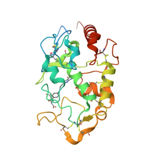
- Additionally, the protein is implicated in neurodegeneration and has a known 3D structure, as indicated by UniProtKB keywords.
Experimental Guidance and Technical Tips
- Consider using a positive control lysate from human kidney or lung tissue, as FOLR1 is highly expressed in these epithelial tissues.
- Validate the antibody in your specific sample system, as expression levels vary between normal and malignant tissues.
- For ELISA, ensure the peptide epitope is accessible under your assay conditions.
- This antibody is reported to cross-react with mouse and rat samples; confirm species cross-reactivity in your experimental setup.
- Optimal working dilutions for WB and ELISA should be determined empirically in your laboratory.
CamelBio: Your One-Stop Sourcing Bridge
CamelBio connects diagnostic manufacturers and research labs with a comprehensive portfolio of IVD raw materials, including validated antibody pairs and optimized monoclonal antibodies like this FOLR1 rabbit mAb. We support your development from concept to clinic with bulk reagents, rare-target sourcing, and technical consultation. Let CamelBio streamline your supply chain for folate receptor-related assays and studies.
Product Datasheet
Folate Binding Protein (FBP) / FOLR1 Rabbit Monoclonal Antibody for WB, ELISA - P15328
REQUEST A QUOTE
Our professional team will reply to you within one business day. Please feel free to contact us!