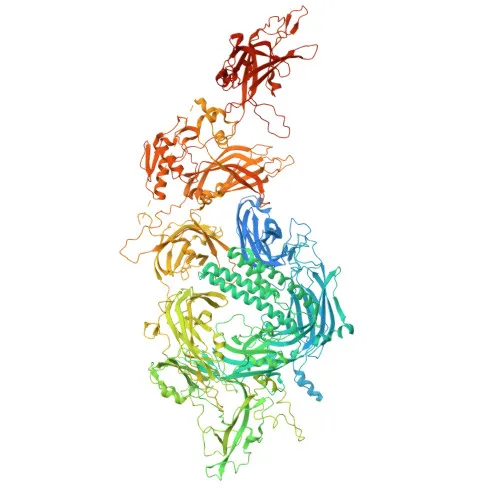
Monoclonal Antibodies
Dysferlin Rabbit Monoclonal Antibody for WB, IHC-P, ELISA - O75923
Item Number : CM0003779
Price varies based on specs and customizations
- Application
- WB, IHC-P, ELISA
- Cross Reactivity
- Human
- Protein Weight
- 237kDa

Shipping:
Contact us to get shipping details Enjoy On-time Dispatch Guarantee.
Why Choose Us
Easy ordering process, quality products, and dedicated support for your business success.
Core Product Specifications and Parameters
| Parameter | Value |
|---|---|
| Product Name | Dysferlin Rabbit mAb |
| Remarks/Alias | MMD1; FER1L1; LGMD2B; LGMDR2; Dysferlin (Romeo) |
| Species | Human |
| GeneID (Human) | 8291 |
| GeneID | 8291 |
| Immunogen | Synthetic Peptide|A synthetic peptide corresponding to a sequence within amino acids 50-150 of human Dysferlin (Romeo) (O75923). |
| Source | Rabbit |
| Category | Monoclonal Antibodies |
| Application | WB, IHC-P, ELISA |
| Cross Reactivity | Human |
| SWISS | O75923 |
| Protein Weight | 237kDa |
| Shipping | Ice bag |
Biological Background: Dysferlin Function and Localization
- Dysferlin (gene DYSF, also known as FER1L1) is a large transmembrane protein belonging to the ferlin family, characterized by multiple C2 domains that mediate calcium-dependent lipid binding.
- It functions as a critical calcium sensor, triggering Ca²⁺-dependent synaptic vesicle–plasma membrane fusion and playing an essential role in the rapid repair of sarcolemmal membranes in skeletal and cardiac muscle following mechanical stress.
- Dysferlin localizes primarily to the cell membrane and sarcolemma, but is also found on cytoplasmic vesicle membranes and late endosomes, consistent with its role in membrane trafficking and repair.
- The protein is heavily expressed in skeletal muscle, heart, brain, spleen, intestine, and placenta, with lower levels in liver, lung, kidney, and pancreas, reflecting its broad tissue distribution.
- Mutations in DYSF are linked to limb-girdle muscular dystrophy type 2B (LGMD2B) and other myopathies, highlighting its clinical significance in neuromuscular disease.
- Dysferlin undergoes alternative promoter usage and alternative splicing, and is subject to phosphorylation; it has been identified in proteomics screens, underscoring its regulatory complexity.
Experimental Guidance and Technical Tips
- The immunogen is a synthetic peptide from the N‑terminal region (aa 50–150) of human Dysferlin; consider epitope mapping if precise binding information is required.
- For Western blotting, use low‑percentage polyacrylamide gels and extended transfer times due to the high molecular weight (237 kDa) of Dysferlin.
- In immunohistochemistry (IHC‑P), validate the antibody on positive control tissues known to express Dysferlin, such as skeletal muscle or heart.
- As a rabbit monoclonal antibody, it is expected to offer high specificity and lot‑to‑lot consistency; however, optimal working dilutions should be determined empirically in the experimenter’s own system.
- For ELISA applications, ensure the peptide immunogen does not interfere with detection if the assay involves the same N‑terminal region.
CamelBio: Your One-Stop Sourcing Bridge
CamelBio serves as a comprehensive sourcing partner for IVD raw materials, supporting diagnostic manufacturers, laboratories, and research institutes from concept to clinic. For targets like Dysferlin—critical in neuromuscular disease research—we offer validated antibody pairs, optimized monoclonal/polyclonal antibodies, and bulk ancillary reagents. Whether you need rare‑target raw‑material sourcing or technical consultation, CamelBio is your trusted one‑stop bridge.
REQUEST A QUOTE
Our professional team will reply to you within one business day. Please feel free to contact us!