Polyclonal Antibodies
CD1B Rabbit pAb for WB, ELISA - P29016
Item Number : CM0030016
Price varies based on specs and customizations
- Application
- WB, ELISA
- Cross Reactivity
- Human
- Protein Weight
- 37kDa

Shipping:
Contact us to get shipping details Enjoy On-time Dispatch Guarantee.
Why Choose Us
Easy ordering process, quality products, and dedicated support for your business success.
Core Product Specifications and Parameters
| Parameter | Value |
|---|---|
| Product Name | CD1B Rabbit pAb |
| Remarks/Alias | R1; CD1; CD1A; CD1B |
| Species | Human |
| Gene ID (Human) | 910 |
| Immunogen | Recombinant fusion protein containing a sequence corresponding to amino acids 18-303 of human CD1B (NP_001755.1) |
| Source | Rabbit |
| Category | Polyclonal Antibodies |
| Application | WB, ELISA |
| Cross Reactivity | Human |
| SWISS | P29016 |
| Protein Weight | 37kDa |
| Shipping | Ice bag |
Biological Background: CD1B Function and Localization
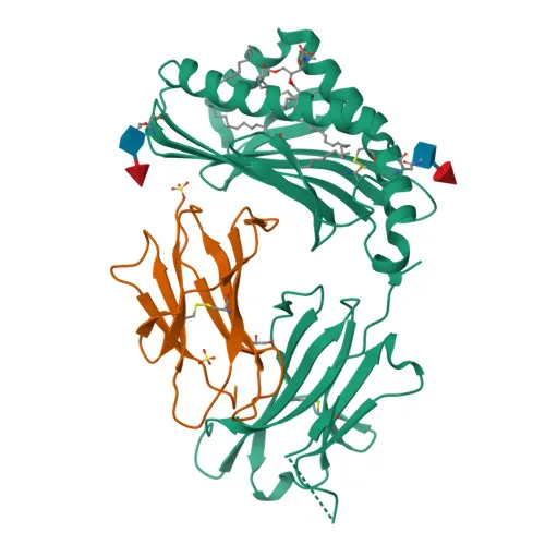
- T-cell surface glycoprotein CD1b is a member of the CD1 family of antigen-presenting molecules, structurally related to MHC class I proteins.
- CD1B specifically binds and presents self and non-self lipid and glycolipid antigens to T-cell receptors on natural killer T (NKT) cells, playing a crucial role in adaptive immunity against microbial pathogens.
- The protein is expressed on the surface of cortical thymocytes, certain T-cell leukemias, and in various other tissues, indicating a role in immune surveillance.
- CD1B localizes to the cell membrane and intracellular compartments such as endosome and lysosome membranes, where it loads lipid antigens in acidic environments.
- It contains an immunoglobulin-like domain, a signal peptide, a transmembrane helix, and short cytoplasmic tail, with key structural features including disulfide bonds and N-linked glycosylation sites.
- Alternative splicing generates multiple isoforms of CD1B, potentially affecting localization and antigen-presenting capacity.
Experimental Guidance and Technical Tips
- The immunogen, consisting of the extracellular domain (aa 18-303), suggests that this antibody may recognize both native and denatured CD1B.
- For Western blot, consider using reducing sample buffer to fully denature the disulfide-bonded immunoglobulin domain; validate in human thymocyte lysates or CD1B-expressing cell lines.
- For ELISA, optimize coating antigen and antibody concentrations using recombinant CD1B protein; the antibody may be suitable for both direct and sandwich formats with appropriate secondary detection.
- Confirm specificity by pre-incubating with immunogen in competition assays, and include proper controls for lipid-presentation studies.
CamelBio: Your One-Stop Sourcing Bridge
CamelBio provides diagnostic manufacturers and research labs with reliable access to IVD raw materials, technical services, and consulting—covering every stage from concept to clinic. This CD1B rabbit polyclonal antibody supports research into lipid antigen presentation and immune profiling. We offer validated antibody pairs, optimized monoclonal and polyclonal antibodies, and bulk ancillary reagents to accelerate your assay development and rare target sourcing needs.
REQUEST A QUOTE
Our professional team will reply to you within one business day. Please feel free to contact us!