Polyclonal Antibodies
BST1 Rabbit Polyclonal Antibody - Q10588
Item Number : CM0025367
Price varies based on specs and customizations
- Application
- WB, IF/ICC, ELISA
- Cross Reactivity
- Human, Mouse
- Protein Weight
- 36kDa

Shipping:
Contact us to get shipping details Enjoy On-time Dispatch Guarantee.
Why Choose Us
Easy ordering process, quality products, and dedicated support for your business success.
Core Product Specifications and Parameters
| Parameter | Value |
|---|---|
| Product Name | BST1 Rabbit pAb |
| Remarks/Alias | CD157; cADPR2; BST1 |
| Species | Human |
| GeneID (Human) | 683 |
| GeneID | 683 |
| Immunogen | Recombinant protein|Recombinant fusion protein containing a sequence corresponding to amino acids 29-293 of human BST1 (NP_004325.2). |
| Source | Rabbit |
| Category | Polyclonal Antibodies |
| Application | WB, IF/ICC, ELISA |
| Cross Reactivity | Human, Mouse |
| SWISS | Q10588 |
| Protein Weight | 36kDa |
| Shipping | Ice bag |
Biological Background: BST1 Function and Localization
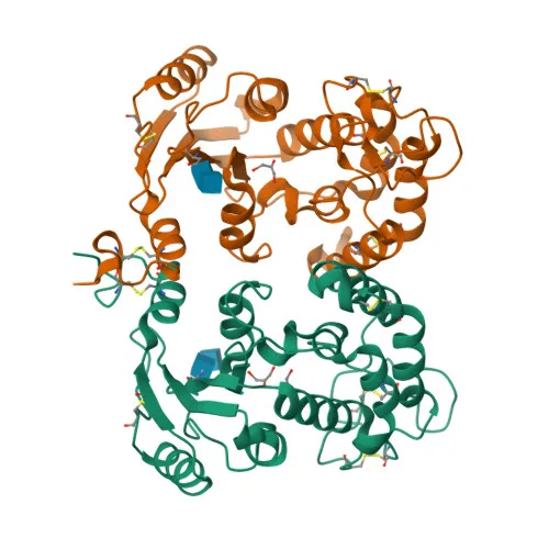
- BST1 encodes ADP-ribosyl cyclase/cyclic ADP-ribose hydrolase 2, which catalyzes both the synthesis of cyclic ADP-ribose (cADPR) from NAD+ and its hydrolysis to ADP-ribose. cADPR serves as a second messenger that elicits calcium release from intracellular stores. Related references: PMID:7805847
- cADPR functions as an endogenous second messenger that regulates the mobilization of intracellular calcium.
- BST1 may be involved in pre-B-cell growth.
- The protein localizes to the cell membrane and is attached via a glycosylphosphatidylinositol (GPI) anchor.
- BST1 is expressed in various tissues including placenta, lung, liver, and kidney.
- Post-translational modifications include glycosylation and disulfide bond formation, which are essential for proper folding and function.
Experimental Guidance and Technical Tips
- The immunogen corresponds to amino acids 29–293 of human BST1, which covers the predicted extracellular domain lacking the GPI-anchor signal. This design may favor recognition of the native protein in non-denaturing applications such as IF/ICC and ELISA.
- For Western blotting, the expected band is approximately 36 kDa under reducing conditions, but glycosylation and other modifications may cause slight mobility shifts. Use appropriate positive controls (e.g., placental or lung lysates) and validate the antibody in your specific sample system.
- For immunofluorescence, consider cell permeabilization if the target epitope is intracellular, or use non-permeabilized conditions for surface staining. BST1 is a GPI-anchored membrane protein, so staining may be predominantly membranous.
- In ELISA, the antibody can be used as a detection reagent after confirming specificity against the recombinant immunogen or native protein. Pairing with a validated capture antibody may improve assay performance.
- Cross-reactivity with mouse BST1 has been reported; however, experimental conditions should be optimized for each species and application.
CamelBio: Your One-Stop Sourcing Bridge
CamelBio provides diagnostic manufacturers, labs, and research institutes with one-stop access to IVD raw materials, technical services, and consulting—covering every stage from concept to clinic. For BST1-related research and assay development, we supply validated antibody pairs, optimized polyclonal antibodies, and bulk ancillary reagents to support your workflow. Our expertise includes sourcing rare-target raw materials, ensuring reliable supply for your diagnostic needs.
REQUEST A QUOTE
Our professional team will reply to you within one business day. Please feel free to contact us!