Polyclonal Antibodies
ASC/TMS1 Rabbit Polyclonal Antibody for WB, IF, IHC-P, ELISA - Q9ULZ3
Item Number : CM0015998
Price varies based on specs and customizations
- Application
- WB, IF/ICC, IF-P, IHC-P, ELISA
- Cross Reactivity
- Human, Mouse, Rat
- Protein Weight
- 22kDa

Shipping:
Contact us to get shipping details Enjoy On-time Dispatch Guarantee.
Why Choose Us
Easy ordering process, quality products, and dedicated support for your business success.
Core Product Specifications and Parameters
| Parameter | Value |
|---|---|
| Product Name | ASC/TMS1 Rabbit pAb |
| Remarks/Alias | ASC; TMS; TMS1; CARD5; TMS-1; ASC/TMS1 |
| Species | Human |
| Gene ID (Human) | 29108 |
| Gene ID | 29108 |
| Immunogen | Synthetic peptide corresponding to amino acids 50-195 of human ASC/TMS1 (NP_037390.2) |
| Source | Rabbit |
| Category | Polyclonal Antibodies |
| Application | WB, IF/ICC, IF-P, IHC-P, ELISA |
| Cross Reactivity | Human, Mouse, Rat |
| SWISS | Q9ULZ3 |
| Protein Weight | 22kDa |
| Shipping | Ice bag |
Biological Background: ASC/TMS1 Function and Localization
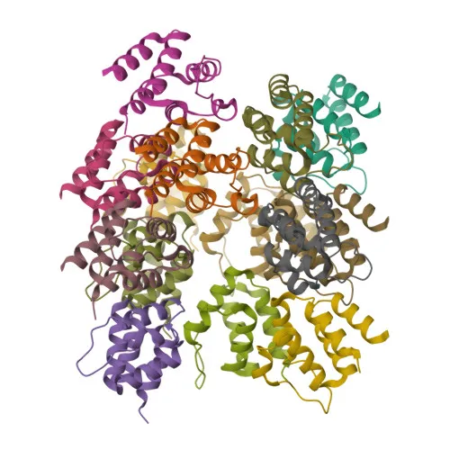
- ASC/TMS1 (apoptosis-associated speck-like protein containing a CARD), also known as PYCARD, CARD5, or TMS-1, is a 22 kDa adapter protein comprising an N-terminal pyrin domain (PYD) and a C-terminal caspase recruitment domain (CARD).
- It acts as a central inflammasome scaffold, bridging NLR sensors (NLRP1, NLRP3, NLRC4) or AIM2 to procaspase-1, thereby driving caspase-1 activation, maturation of IL-1β and IL-18, and pyroptotic cell death. Related references: PMID:15030775, PMID:16982856, PMID:17349957, PMID:24630722
- ASC mediates apoptosis by promoting caspase-8-dependent cleavage of BID, mitochondrial BAX translocation, and subsequent caspase-9 and -3 activation, often independently of FADD. Related references: PMID:11103777, PMID:12646168, PMID:14730312, PMID:16964285
- In the nucleus, ASC can modulate NF-κB signaling by inhibiting the IKK complex and competing with RIPK2, thereby finetuning inflammatory gene expression. Related references: PMID:12486103, PMID:16585594
- ASC localizes to multiple subcellular compartments, including cytoplasm, inflammasome specks, endoplasmic reticulum, mitochondria, nucleus, and Golgi, consistent with its versatile roles in immune signaling and apoptosis.
- Widely expressed at low levels, with higher abundance in peripheral blood leukocytes, lung, small intestine, spleen, thymus, and colon. Expression is often silenced by promoter methylation in tumors, aligning with its reported tumor suppressor activity. Related references: PMID:23229815
- ASC is subject to regulatory post-translational modifications, including ubiquitination and phosphorylation, which control inflammasome assembly and signal strength.
Experimental Guidance and Technical Tips
- The immunogen spans the central region (aa 50–195) bridging the PYD and CARD domains, which may confer reactivity against both full-length ASC and splice variants; consider validating specificity by knockdown or knockout controls.
- In Western blot, a predominant band around 22 kDa is expected; use positive control lysates from THP‑1 cells or human peripheral blood mononuclear cells, and include protease inhibitors to minimize degradation.
- For immunofluorescence and ICC, speck‑like aggregates are a hallmark of activated inflammasomes; co‑staining with antibodies against NLRP3, AIM2, or caspase‑1 can confirm inflammasome formation. Fixation and permeabilization conditions should be optimized for the cell type and activation state.
- In IHC‑P (paraffin‑embedded tissues), antigen retrieval in citrate buffer (pH 6.0) and blocking endogenous peroxidases are recommended; tissues with known inflammation (e.g., inflamed colon, tonsil) serve as suitable positive controls.
- For ELISA applications, pairing this polyclonal antibody with a capture monoclonal antibody may improve signal; validate assay linearity and specificity with recombinant ASC protein.
- This antibody detects ASC from human, mouse, and rat; however, cross‑reactivity may vary by assay format, so it is advised to include species‑specific validation.
CamelBio: Your One-Stop Sourcing Bridge
CamelBio streamlines IVD raw material procurement for diagnostic manufacturers, academic labs, and contract research organizations. Our portfolio includes validated antibody pairs, recombinant antigens, and bulk ancillary reagents, all backed by technical consultation from target selection to clinical validation. For ASC/TMS1 and other inflammasome‑related targets, we offer high‑quality polyclonal and monoclonal antibodies, and we can source custom formulations to support your unique assay development needs.
Product Datasheet
ASC/TMS1 Rabbit Polyclonal Antibody for WB, IF, IHC-P, ELISA - Q9ULZ3
REQUEST A QUOTE
Our professional team will reply to you within one business day. Please feel free to contact us!