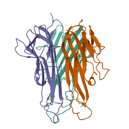
Monoclonal Antibodies
APC Rabbit anti-Mouse TNF-α Monoclonal Antibody for FC (intra) - P06804
Item Number : CM0011495
Price varies based on specs and customizations
- Application
- FC (intra)
- Cross Reactivity
- Mouse
- Protein Weight
- 26kDa

Shipping:
Contact us to get shipping details Enjoy On-time Dispatch Guarantee.
Why Choose Us
Easy ordering process, quality products, and dedicated support for your business success.
Core Product Specifications and Parameters
| Parameter | Value |
|---|---|
| Product Name | APC Rabbit anti-Mouse TNF-α mAb |
| Remarks/Alias | DIF; Tnfa; TNF-a; TNFSF2; Tnlg1f; Tnfsf1a; TNFalpha; TNF-alpha |
| Species | Human |
| GeneID (Human) | 7124 |
| GeneID | 21926 |
| Immunogen | Recombinant fusion protein containing a sequence corresponding to amino acids 80-235 of mouse TNF-α (NP_038721.1) |
| Source | Rabbit |
| Category | Monoclonal Antibodies |
| Application | FC (intra) |
| Cross Reactivity | Mouse |
| SWISS | P06804 |
| Protein Weight | 26kDa |
| Shipping | Ice bag |
Biological Background: TNF-α Function and Localization
- Tumor necrosis factor (TNF-α) is a pro-inflammatory cytokine primarily secreted by activated macrophages, belonging to the tumor necrosis factor ligand superfamily (TNFSF2). It is also known as cachectin and is encoded by the Tnf gene (alternative names Tnfa, Tnfsf2).
- TNF-α exerts its biological effects through binding to two distinct receptors, TNFR1 (TNFRSF1A) and TNFR2 (TNFRSF1B), mediating a wide range of cellular responses including apoptosis, cell survival, and inflammation.
- This cytokine is a key mediator of the acute phase response and can function as an endogenous pyrogen, inducing fever directly or through stimulation of interleukin-1 secretion, and is implicated in the development of cachexia in chronic disease states.
- In adipose tissue, TNF-α induces insulin resistance by inhibiting insulin-induced IRS1 tyrosine phosphorylation and glucose uptake, and promotes degradation of GKAP42 protein, contributing to the pathogenesis of type 2 diabetes. Related references: PMID:25586176
- TNF-α plays a significant role in bone metabolism by promoting osteoclastogenesis and thereby mediating bone resorption, linking inflammation to bone loss in conditions such as rheumatoid arthritis. Related references: PMID:32741026
- The intracellular domain (ICD) of TNF-α, generated by proteolytic cleavage, can act as a signaling molecule to induce IL-12 production in dendritic cells, highlighting a receptor-independent function.
- Subcellularly, TNF-α is synthesized as a type II transmembrane protein (26 kDa) on the cell membrane, and is cleaved by TNF-α converting enzyme (TACE) to release the soluble 17 kDa cytokine. The protein undergoes several post-translational modifications including glycosylation, phosphorylation, myristoylation, and disulfide bond formation.
- Besides its cytotoxic and inflammatory roles, TNF-α also stimulates angiogenesis synergistically with IL-1β and IL-6, and under certain conditions can promote cell proliferation and differentiation, reflecting its pleiotropic nature.
Experimental Guidance and Technical Tips
- The immunogen corresponds to amino acids 80-235 of mouse TNF-α (NP_038721.1), covering the mature soluble TNF-α region. This design targets the extracellular domain, potentially recognizing both the membrane-bound and soluble forms.
- For intracellular flow cytometry, fixation and permeabilization protocols are critical. Consider using a standard intracellular staining protocol with a fixation/permeabilization kit suitable for cytokines.
- Since TNF-α is a secreted protein, resting cells may have low intracellular levels. For robust detection, stimulate cells (e.g., with PMA/ionomycin or LPS) in the presence of a protein transport inhibitor (e.g., Brefeldin A or Monensin) to accumulate TNF-α in the Golgi apparatus.
- It is recommended to titrate the antibody to determine the optimal concentration for your specific experimental system, starting with a range of 0.5-5 µg per million cells.
- Run appropriate controls including unstimulated cells and isotype control antibodies to differentiate specific staining from background.
- The antibody is conjugated to APC, a bright fluorophore suitable for multicolor flow cytometry panels. Ensure proper compensation when used with other fluorophores.
CamelBio: Your One-Stop Sourcing Bridge
CamelBio serves as a comprehensive sourcing partner for IVD raw materials, supporting diagnostic manufacturers, laboratories, and research institutes from concept to clinic. This APC-conjugated anti-mouse TNF-α antibody is well-suited for developing sensitive flow cytometry-based assays and immunoassay platforms, and CamelBio can assist with bulk procurement, customized conjugations, and complementary reagent sourcing. With reliable access to validated antibody pairs, optimized monoclonal/polyclonal antibodies, and ancillary reagents, CamelBio simplifies your supply chain for inflammation and immunology research.
Product Datasheet
APC Rabbit anti-Mouse TNF-α Monoclonal Antibody for FC (intra) - P06804
REQUEST A QUOTE
Our professional team will reply to you within one business day. Please feel free to contact us!