Monoclonal Antibodies
APC Rabbit anti-Mouse CD317/BST2 Monoclonal Antibody for FC - Q8R2Q8
Item Number : CM0007731
Price varies based on specs and customizations
- Application
- FC
- Cross Reactivity
- Mouse
- Protein Weight
- 19kDa

Shipping:
Contact us to get shipping details Enjoy On-time Dispatch Guarantee.
Why Choose Us
Easy ordering process, quality products, and dedicated support for your business success.
Core Product Specifications and Parameters
| Parameter | Value |
|---|---|
| Product Name | APC Rabbit anti-Mouse CD317/BST2 mAb |
| Remarks/Alias | GREG; Bst-2; CD317; DAMP-1; 2310015I10Rik |
| Species | Mouse |
| Gene ID (Human) | 684 |
| Gene ID | 69550 |
| Immunogen | Recombinant fusion protein containing a sequence corresponding to amino acids 55-152 of mouse CD317/BST2 (NP_932763.1). |
| Source | Rabbit |
| Category | Monoclonal Antibodies |
| Application | FC |
| Cross Reactivity | Mouse |
| SWISS | Q8R2Q8 |
| Protein Weight | 19kDa |
| Shipping | Ice bag |
Biological Background: CD317/BST2 Function and Localization
- CD317/BST2 (bone marrow stromal antigen 2), also known as HM1.24 antigen, is a 19 kDa protein encoded by the Bst2 gene in mouse (Gene ID: 69550).
- It functions as an interferon-induced antiviral host restriction factor, blocking release of diverse enveloped viruses (e.g., HIV-1, MMTV, MLV, EBOV, LASV, VSV) by directly tethering nascent virions to cell membranes.
- The protein acts as a physical tether, linking virions to the cell surface and to each other, leading to virus internalization and degradation or surface retention, thus restricting cell-free spread.
- CD317/BST2 also regulates cell growth and migration by inhibiting cell surface proteolytic activity of MMP14, which in turn decreases MMP15 activation.
- In plasmacytoid dendritic cells, it stimulates LILRA4/ILT7 signaling, providing negative feedback to type I interferon production during viral infection.
- It plays a role in organizing the subapical actin cytoskeleton in polarized epithelial cells.
- Subcellularly, CD317/BST2 localizes to the Golgi apparatus, trans-Golgi network, cell membrane, late endosomes, membrane rafts, cytoplasm, and apical cell membrane.
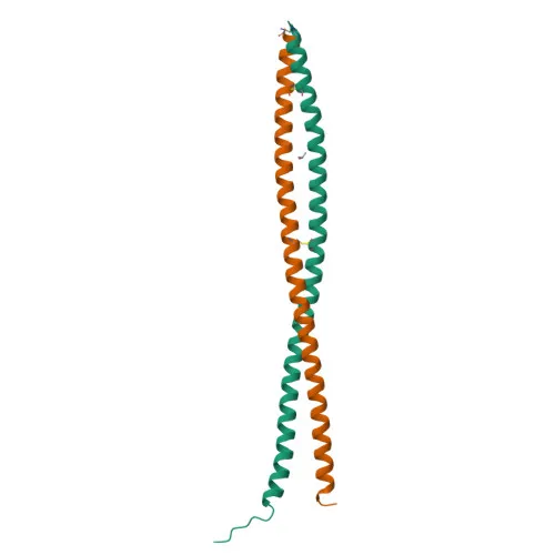
- The protein is specifically expressed on type I interferon-producing cells in naive mice, and features a GPI-anchor, coiled coil domains, disulfide bonds, and glycosylation.
Experimental Guidance and Technical Tips
- This APC-conjugated antibody is designed for flow cytometry; consider direct detection of surface CD317 on live mouse cells without secondary reagents.
- Validate staining in relevant sample systems, such as plasmacytoid dendritic cells or interferon-treated cell lines, as CD317 expression is IFN-inducible.
- For intracellular staining, include appropriate permeabilization steps, as CD317 localizes to multiple membrane compartments.
- When co-staining with other fluorophores, ensure compensation controls, as APC signal can be bright.
CamelBio: Your One-Stop Sourcing Bridge
CamelBio streamlines IVD raw-material procurement by offering validated antibody pairs, optimized monoclonal/polyclonal antibodies, and bulk ancillary reagents. This anti-CD317/BST2 antibody supports antiviral innate immunity and dendritic cell research, aligning with CamelBio's commitment to supplying rare-target reagents. Partner with CamelBio for reliable access to critical raw materials, from concept to clinic.
REQUEST A QUOTE
Our professional team will reply to you within one business day. Please feel free to contact us!