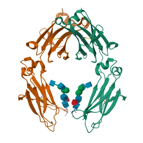
Monoclonal Antibodies
APC Rabbit anti-Human IgG (Fc) Monoclonal Antibody for Flow Cytometry - P01857
Item Number : CM0009241
Price varies based on specs and customizations
- Application
- FC
- Cross Reactivity
- Human
- Protein Weight
- 36 kDa

Shipping:
Contact us to get shipping details Enjoy On-time Dispatch Guarantee.
Why Choose Us
Easy ordering process, quality products, and dedicated support for your business success.
Core Product Specifications and Parameters
| Parameter | Value |
|---|---|
| Product Name | APC Rabbit anti-Human IgG (Fc) mAb |
| Remarks/Alias | IGHG1 |
| Species | Human |
| Gene ID (Human) | 3500 |
| Gene ID | 3500 |
| Immunogen | Recombinant protein (or fragment). This information is considered to be commercially sensitive. |
| Source | Rabbit |
| Category | Monoclonal Antibodies |
| Application | FC |
| Cross Reactivity | Human |
| SWISS | P01857 |
| Protein Weight | 36 kDa |
| Shipping | Ice bag |
Biological Background: Human IgG1 Constant Region Function and Localization
- Immunoglobulin heavy constant gamma 1 (IGHG1) is the constant region of the gamma 1 heavy chain, defining the IgG1 isotype. It is expressed as both a membrane-bound receptor on B cells and a secreted antibody in plasma.
- Membrane-bound IgG1 serves as the B-cell antigen receptor, triggering clonal expansion and differentiation upon antigen binding. Secreted IgG1 antibodies mediate the effector phase of humoral immunity via their constant (Fc) region.
- The antigen-binding region is formed by the variable domain of the heavy chain paired with the light chain. Remarkable antigen affinity is achieved through V-(D)-J rearrangement, somatic hypermutation, and affinity maturation.
- The constant region of IgG1 interacts via its Fc portion with Fc receptors on innate immune cells and with the complement system. This interaction mediates antibody-dependent cellular cytotoxicity (ADCC), complement-dependent cytotoxicity (CDC), and inflammatory responses, leading to pathogen neutralization. Related references: PMID:17576170 PMID:20176268 PMID:22158414
- Subcellularly, IgG1 is found secreted and anchored to the cell membrane of B lymphocytes. The membrane-bound form contains a transmembrane helix.
- Key features include disulfide bonds that stabilize the immunoglobulin fold, glycosylation sites important for effector functions, and the capacity for alternative splicing. The generation of IgG1 involves class-switch recombination from IgM/IgD, diversifying the antibody repertoire.
Experimental Guidance and Technical Tips
- The immunogen corresponds to a recombinant fragment of the Fc region; therefore the antibody reacts with the constant region of human IgG1 heavy chains. It is expected to recognize both membrane and secreted forms.
- This conjugate is validated for flow cytometry. For optimal results, titrate the antibody on the target cell population to determine the saturation concentration and minimize background. Include appropriate isotype controls.
- As an APC-conjugated reagent, protect the antibody from prolonged light exposure and avoid multiple freeze–thaw cycles. Store according to the manufacturer’s instructions.
- In flow cytometry, consider using a viability dye to exclude dead cells, as they may bind antibodies non‑specifically. Confirm specificity by staining cells with known IgG expression profiles.
CamelBio: Your One‑Stop Sourcing Bridge
For diagnostic manufacturers, labs, and research institutes, CamelBio serves as a comprehensive IVD raw‑material partner, from concept to clinic. This anti‑human IgG (Fc) antibody supports immunology, B‑cell biology, and therapeutic antibody development workflows. Beyond single reagents, CamelBio offers validated antibody pairs, optimized monoclonals and polyclonals, bulk ancillary reagents, and rare‑target sourcing — all backed by expert technical consultation to streamline your assay development.
Product Datasheet
APC Rabbit anti-Human IgG (Fc) Monoclonal Antibody for Flow Cytometry - P01857
REQUEST A QUOTE
Our professional team will reply to you within one business day. Please feel free to contact us!