Monoclonal Antibodies
APC Anti-Human TLR3 Monoclonal Antibody for Flow Cytometry - O15455
Item Number : CM0011928
Price varies based on specs and customizations
- Application
- FC (intra)
- Cross Reactivity
- Human
- Protein Weight
- 104kDa

Shipping:
Contact us to get shipping details Enjoy On-time Dispatch Guarantee.
Why Choose Us
Easy ordering process, quality products, and dedicated support for your business success.
Core Product Specifications and Parameters
| Parameter | Value |
|---|---|
| Product Name | APC Rabbit anti-Human TLR3 mAb |
| Remarks/Alias | CD283; IIAE2; IMD83 |
| Species | Human |
| GeneID (Human) | 7098 |
| GeneID | 7098 |
| Immunogen | Recombinant protein corresponding to a sequence within amino acids 24-339 of human TLR3 (NP_003256.1) |
| Source | Rabbit |
| Category | Monoclonal Antibodies |
| Application | FC (intra) |
| Cross Reactivity | Human |
| SWISS | O15455 |
| Protein Weight | 104kDa |
| Shipping | Ice bag |
Biological Background: TLR3 Function and Localization
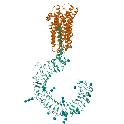
- Toll-like receptor 3 (TLR3), also known as CD283, IIAE2, and IMD83, is a single-pass type I membrane protein that belongs to the Toll-like receptor family characterized by leucine-rich repeats (LRRs) in the extracellular domain and a Toll/interleukin-1 receptor (TIR) domain in the cytoplasmic tail.
- TLR3 is a key component of both innate and adaptive immunity, functioning as a pattern recognition receptor that senses double-stranded RNA (dsRNA), a molecular hallmark of viral infection, thereby triggering antiviral immune responses.
- Upon dsRNA binding in endosomes, TLR3 signals exclusively through the adaptor protein TRIF (TICAM1), which activates transcription factors NF-kappa-B and IRF3, leading to the production of type I interferons and proinflammatory cytokines.
- Subcellularly, TLR3 localizes primarily to the endoplasmic reticulum membrane, endosome membrane, and early endosomes, positioning it to detect viral dsRNA released during endosomal acidification.
- TLR3 is highly expressed in the placenta and pancreas, and is prominently found in CD11c+ immature dendritic cells; notably, it is the most strongly expressed TLR in the brain, particularly in astrocytes, glia, and neurons.
- The protein undergoes post-translational modifications including glycosylation, phosphorylation, and ubiquitin conjugation, which regulate its trafficking and signaling activity.
- Alternative splicing generates multiple isoforms, and mutations in the TLR3 gene are associated with immunodeficiency and susceptibility to herpes simplex encephalitis.
- With a molecular weight of approximately 104 kDa, TLR3 contains multiple disulfide bonds that stabilize its solenoid-shaped LRR ectodomain, essential for ligand recognition.
Experimental Guidance and Technical Tips
- The immunogen corresponds to the N-terminal extracellular region (amino acids 24–339), which encompasses several LRR modules; ensure the antibody recognizes the native conformation if used for cell surface or endosomal staining under non-denaturing conditions.
- For intracellular flow cytometry (FC), permeabilization is required; test fixation and permeabilization protocols (e.g., saponin or Triton X-100) to optimize epitope exposure while preserving fluorophore stability.
- TLR3 expression is predominantly intracellular in resting cells; stimulation with dsRNA or viral infection may upregulate and redistribute the receptor, so consider appropriate positive controls such as poly(I:C)-treated PBMCs or dendritic cells.
- Cross-reactivity with other species is not reported; validate detection in the intended sample matrix, particularly given high TLR3 expression in placenta and brain tissues.
- The APC conjugate provides a bright signal for flow cytometry; use appropriate compensation controls and avoid prolonged light exposure to prevent photobleaching.
CamelBio: Your One-Stop Sourcing Bridge
CamelBio streamlines the procurement of IVD raw materials, offering a comprehensive portfolio of validated antibodies, recombinant proteins, and ancillary reagents. Supporting research in innate immunity and viral pathogenesis, this anti-TLR3 antibody is part of CamelBio’s extensive catalog designed to accelerate assay development and diagnostic manufacturing. Through a global supply network and technical consultation, CamelBio ensures efficient access to specialized raw materials, from early-stage discovery to clinical application.
REQUEST A QUOTE
Our professional team will reply to you within one business day. Please feel free to contact us!