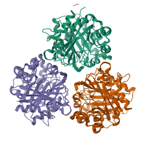
Monoclonal Antibodies
Anti-YKL-40/CHI3L1 Rabbit Monoclonal Antibody for WB, IP, IHC-P, ELISA - Q61362
Item Number : CM0011075
Price varies based on specs and customizations
- Application
- WB, IP, IHC-P, ELISA
- Cross Reactivity
- Mouse
- Protein Weight
- 43 kDa/44 kDa

Shipping:
Contact us to get shipping details Enjoy On-time Dispatch Guarantee.
Why Choose Us
Easy ordering process, quality products, and dedicated support for your business success.
Core Product Specifications and Parameters
| Parameter | Value |
|---|---|
| Product Name | YKL-40/CHI3L1 Rabbit mAb |
| Remarks/Alias | Gp39; Brp39; Chil1 |
| Species | Mouse |
| GeneID (Human) | 1116 |
| GeneID (Mouse) | 12654 |
| Immunogen | Recombinant fusion protein containing a sequence corresponding to amino acids 30-389 of Mouse Chi3l1 (NP_031721.2). |
| Source | Rabbit |
| Category | Monoclonal Antibodies |
| Application | WB, IP, IHC-P, ELISA |
| Cross Reactivity | Mouse |
| SWISS | Q61362 |
| Protein Weight | 43 kDa/44 kDa |
| Shipping | Ice bag |
Biological Background: YKL-40/CHI3L1 Function and Localization
- Encodes multiple isoforms via alternative initiation and alternative splicing.
- Lacks chitinase activity but functions as a carbohydrate-binding lectin with preference for chitin; involved in tissue remodeling and cellular adaptation to environmental changes.
- Plays a key role in Th2 inflammatory responses and IL-13-induced inflammation, regulating allergen sensitization, inflammatory cell apoptosis, dendritic cell accumulation, and M2 macrophage differentiation.
- Facilitates invasion of pathogenic enteric bacteria into colonic mucosa and lymphoid organs; mediates AKT1 signaling and IL8 production in colonic epithelial cells.
- In lung, regulates antibacterial responses by enhancing macrophage bacterial killing, controlling bacterial dissemination, and augmenting host tolerance; also regulates hyperoxia-induced lung injury, inflammation, and epithelial apoptosis.
- Subcellular localization includes secretion into extracellular space, as well as presence in cytoplasm and endoplasmic reticulum.
- Tissue specificity: detected in lung (pulmonary macrophages, alveolar type 2 cells, bronchoalveolar lavage fluid), and in mammary tumor cells; not detected in non-inflammatory colon.
- Annotated keywords: 3D-structure, Alternative initiation, Alternative splicing, Antimicrobial, Apoptosis, Cytoplasm, Disulfide bond, Endoplasmic reticulum, Glycoprotein, Inflammatory response, Reference proteome, Secreted, Signal.
Experimental Guidance and Technical Tips
- Based on the immunogen design spanning amino acids 30-389 of mouse Chi3l1, this antibody may recognize multiple isoforms and is potentially suitable for detecting both full-length and alternative splice variants; consider sequence alignment for cross-reactivity with other species.
- For WB: predicted molecular weight around 43-44 kDa under reducing conditions, but may migrate differently due to glycosylation or alternative initiation; include positive controls such as lung tissue lysates or macrophage cell lines.
- For IHC-P: antigen retrieval may be required; validate in tissues with known expression (e.g., inflamed lung, mammary tumor) and include negative control (non-inflammatory colon).
- For IP and ELISA: ensure buffer compatibility; consider using antibody at recommended concentrations and validate in the relevant sample system.
- Cross-reactivity is reported for mouse; for other species, verify sequence homology and perform appropriate controls.
CamelBio: Your One-Stop Sourcing Bridge
This YKL-40/CHI3L1 antibody supports research in inflammation, tissue remodeling, and host-pathogen interactions. As a specialized IVD raw-material partner, CamelBio offers validated antibody pairs, optimized monoclonal/polyclonal antibodies, bulk ancillary reagents, and efficient sourcing for rare targets to help streamline your diagnostic development from concept to clinic.
Product Datasheet
Anti-YKL-40/CHI3L1 Rabbit Monoclonal Antibody for WB, IP, IHC-P, ELISA - Q61362
REQUEST A QUOTE
Our professional team will reply to you within one business day. Please feel free to contact us!