Polyclonal Antibodies
Anti-WDR1 Polyclonal Antibody for WB and ELISA - O75083
Item Number : CM0015758
Price varies based on specs and customizations
- Application
- WB, ELISA
- Cross Reactivity
- Human, Mouse, Rat
- Protein Weight
- 66kDa

Shipping:
Contact us to get shipping details Enjoy On-time Dispatch Guarantee.
Why Choose Us
Easy ordering process, quality products, and dedicated support for your business success.
Core Product Specifications and Parameters
| Parameter | Value |
|---|---|
| Product Name | WDR1 Rabbit pAb |
| Remarks/Alias | AIP1; PFITS; NORI-1; HEL-S-52; WDR1 |
| Species | Human |
| Gene ID (Human) | 9948 |
| Immunogen | Recombinant protein|Recombinant fusion protein containing a sequence corresponding to amino acids 300-500 of human WDR1 (NP_059830.1) |
| Source | Rabbit |
| Category | Polyclonal Antibodies |
| Application | WB, ELISA |
| Cross Reactivity | Human, Mouse, Rat |
| SWISS | O75083 |
| Protein Weight | 66kDa |
| Shipping | Ice bag |
Biological Background: WDR1 Function and Localization
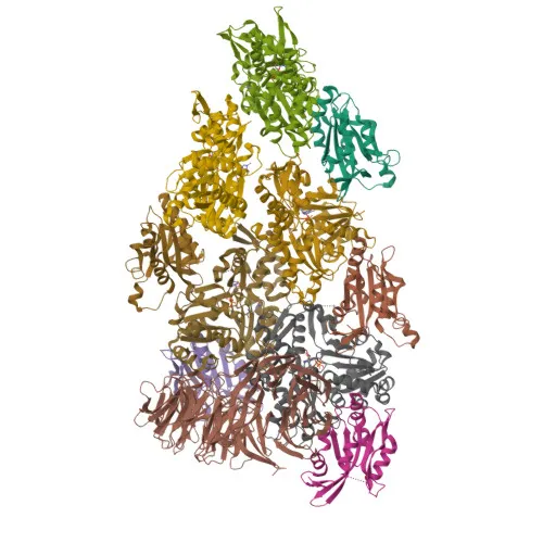
- WDR1 (Actin-interacting protein 1, AIP1, NORI-1) is encoded by the WDR1 gene and plays a central role in actin cytoskeleton remodeling.
- Induces disassembly of actin filaments in conjunction with ADF/cofilin family proteins and enhances cofilin-mediated actin severing. Related references: PMID:15629458 PMID:27557945 PMID:29751004
- Involved in cytokinesis, chemotactic cell migration by restricting lamellipodial protrusions, and is required for cardiomyocyte growth and maintenance (by similarity). Related references: PMID:18494608
- Subcellular localization includes cytoplasm, cytoskeleton, cell projections (podosomes), and cell junctions, where it organizes the perijunctional actomyosin belt. Related references: PMID:25792565
- Plays a role in megakaryocyte maturation, platelet shedding, and establishment of planar cell polarity (by similarity).
- Post-translational modifications include acetylation and phosphorylation, as identified by proteomics.
- Expressed in peripheral blood mononuclear cells at the protein level.
Experimental Guidance and Technical Tips
- The immunogen (amino acids 300-500) targets the central WD repeat region, likely generating antibodies that recognize native WDR1. Confirm specificity by Western blot with a single band at ~66 kDa.
- For Western blot (WB), use standard blocking conditions (e.g., 5% non-fat milk) and validate detection in human, mouse, or rat cell lysates known to express WDR1.
- In ELISA applications, the recombinant immunogen can serve as a positive control; optimize coating conditions and antibody dilution for best signal-to-noise ratio.
- Since this is a polyclonal antibody, lot-to-lot consistency may vary; include a positive control and consider pre-absorption assays for critical applications.
CamelBio: Your One-Stop Sourcing Bridge
CamelBio offers diagnostic manufacturers and research labs streamlined access to validated IVD raw materials, including polyclonal and monoclonal antibodies, recombinant proteins, and ancillary reagents. With expertise in actin cytoskeleton targets like WDR1, we can support your development of assays for cell migration, cytokinesis, or cytoskeletal disease biomarkers. Whether you need established products or custom rare-target sourcing, our one-stop platform covers every stage from concept to clinic.
REQUEST A QUOTE
Our professional team will reply to you within one business day. Please feel free to contact us!