Polyclonal Antibodies
Anti-VTI1B Rabbit Polyclonal Antibody for WB, IHC-P, ELISA - Q9UEU0
Item Number : CM0027521
Price varies based on specs and customizations
- Application
- WB, IHC-P, ELISA
- Cross Reactivity
- Human, Mouse
- Protein Weight
- 27kDa

Shipping:
Contact us to get shipping details Enjoy On-time Dispatch Guarantee.
Why Choose Us
Easy ordering process, quality products, and dedicated support for your business success.
Core Product Specifications and Parameters
| Parameter | Value |
|---|---|
| Product Name | VTI1B Rabbit pAb |
| Remarks/Alias | VTI1; VTI2; VTI1L; v-SNARE; vti1-rp1; VTI1-LIKE; VTI1B |
| Species | Human |
| GeneID | 10490 |
| Immunogen | Recombinant fusion protein containing a sequence corresponding to amino acids 1-210 of human VTI1B (NP_006361.1). |
| Source | Rabbit |
| Category | Polyclonal Antibodies |
| Application | WB, IHC-P, ELISA |
| Cross Reactivity | Human, Mouse |
| SWISS | Q9UEU0 |
| Protein Weight | 27kDa |
| Shipping | Ice bag |
Biological Background: VTI1B Function and Localization
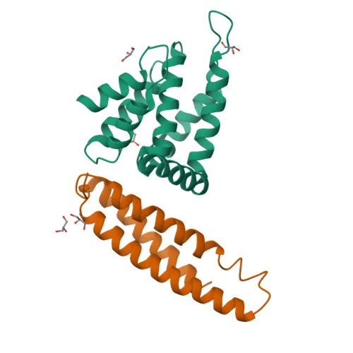
- VTI1B, also known as vesicle transport through interaction with t-SNAREs homolog 1B (Vti1-rp1, VTI1L), is a SNARE protein that functions as a v-SNARE in intracellular membrane fusion.
- It participates in the formation of trans-SNARE complexes, where four-helix bundles assemble to drive membrane fusion, a critical process for vesicular trafficking. Related references: PMID:21388490, PMID:23217709
- Specifically, on late endosomes, VTI1B regulates trafficking from late endosomes to the lysosome, facilitating cargo delivery for degradation.
- The protein is localized to membranes of early endosomes, late endosomes, lysosomes, recycling endosomes, and cytoplasmic granules, highlighting its dynamic role in endosomal sorting.
- VTI1B is a transmembrane protein with a molecular weight of approximately 27 kDa and undergoes post-translational modifications including acetylation, methylation, and phosphorylation.
- It is broadly expressed across all human tissues, reflecting its fundamental role in membrane trafficking pathways.
- Alternative splicing of the VTI1B gene generates multiple isoforms, contributing to functional diversity.
Experimental Guidance and Technical Tips
- The immunogen corresponds to the N-terminal region (amino acids 1-210) of human VTI1B, which is likely to recognize both full-length and truncated isoforms; validate specificity in your sample system.
- For Western blot (WB), the predicted molecular weight is ~27 kDa; consider using a suitable blocking buffer and optimizing primary antibody dilution based on target abundance.
- For immunohistochemistry (IHC-P), antigen retrieval methods may enhance detection; confirm with positive and negative control tissues.
- In ELISA, the polyclonal nature may provide high sensitivity; quantify using purified VTI1B protein or peptide as standard to determine antibody titer.
- The antibody cross-reacts with human and mouse VTI1B; if using in other species, verify sequence homology and perform validation experiments.
- Since VTI1B is a membrane protein, inclusion of mild detergents in lysis buffers may improve solubilization and detection.
CamelBio: Your One-Stop Sourcing Bridge
CamelBio is your trusted partner for IVD raw-material procurement, offering validated antibodies, recombinant proteins, and ancillary reagents for diagnostic and research applications. This VTI1B rabbit polyclonal antibody is part of our extensive catalog supporting SNARE trafficking and endosomal research, and we can also source custom formulations or bulk quantities to meet your specific assay development needs. Contact us to streamline your supply chain from concept to clinic.
REQUEST A QUOTE
Our professional team will reply to you within one business day. Please feel free to contact us!